. 2009 Jul;85(1):76-86.
doi: 10.1016/j.ajhg.2009.06.006. Epub 2009 Jul 2.

Deficiency of Dol-P-Man synthase subunit DPM3 bridges the congenital disorders of glycosylation with the dystroglycanopathies

Affiliations

Deficiency of Dol-P-Man synthase subunit DPM3 bridges the congenital disorders of glycosylation with the dystroglycanopathies

Dirk J Lefeber et al. Am J Hum Genet. 2009 Jul.

Abstract

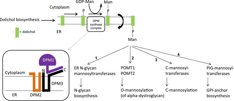
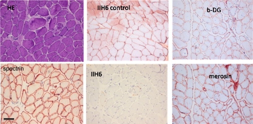
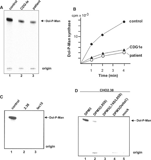
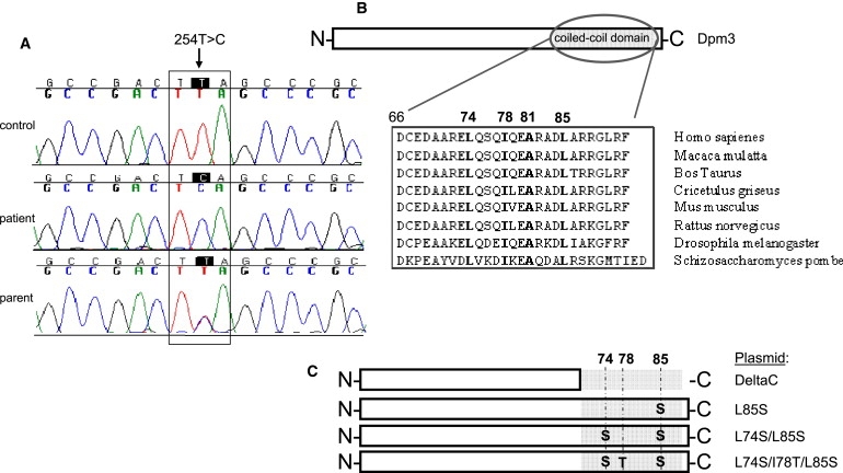
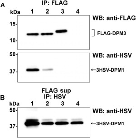
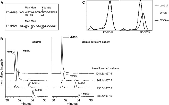
Alpha-dystroglycanopathies such as Walker Warburg syndrome represent an important subgroup of the muscular dystrophies that have been related to defective O-mannosylation of alpha-dystroglycan. In many patients, the underlying genetic etiology remains unsolved. Isolated muscular dystrophy has not been described in the congenital disorders of glycosylation (CDG) caused by N-linked protein glycosylation defects. Here, we present a genetic N-glycosylation disorder with muscular dystrophy in the group of CDG type I. Extensive biochemical investigations revealed a strongly reduced dolichol-phosphate-mannose (Dol-P-Man) synthase activity. Sequencing of the three DPM subunits and complementation of DPM3-deficient CHO2.38 cells showed a pathogenic p.L85S missense mutation in the strongly conserved coiled-coil domain of DPM3 that tethers catalytic DPM1 to the ER membrane. Cotransfection experiments in CHO cells showed a reduced binding capacity of DPM3(L85S) for DPM1. Investigation of the four Dol-P-Man-dependent glycosylation pathways in the ER revealed strongly reduced O-mannosylation of alpha-dystroglycan in a muscle biopsy, thereby explaining the clinical phenotype of muscular dystrophy. This mild Dol-P-Man biosynthesis defect due to DPM3 mutations is a cause for alpha-dystroglycanopathy, thereby bridging the congenital disorders of glycosylation with the dystroglycanopathies.


Figures

Figure 1
Figure 1
Architecture of the Dol-P-Man Synthase Complex The enzymatically active DPM1 subunit in the cytoplasm is anchored to subunits DPM2 and DPM3 in the ER membrane via the C-terminal peptide of DPM3. Four biosynthetic pathways depend on Dol-P-Man: N-glycosylation (1), O-mannosylation (2), C-Mannosylation (3), and GPI-anchor biosynthesis (4).
Figure 2
Figure 2
Muscle Biopsy of m. rectus femoris at 20 Years Histochemistry with hematoxylin-phloxin (HE) and immunohistochemistry with IIH6 antibody against glycosylated alpha-dystroglycan and with antibodies against spectrin, merosin, and β-dystroglycan (b-DG). A staining of a control muscle biopsy with IIH6 is shown for comparison. The scale bar represents 100 μm.
Figure 3
Figure 3
Determination of CDG Subtype (A) Isoelectric focusing of serum transferrin. Profiles of patient and both parents are shown in lanes 3 and 4 + 5, respectively. Controls are presented in lane 1 (healthy control), lane 2 (CDG-Ia), lane 6 (CDG-II patient), and lane 7 (CDG-Ie). Numbers 0, 2, 3, and 4 indicate the sialotransferrin subfractions. (B) Maldi mass spectrometry of permethylated N-glycans from serum expressed as relative percentage of the biantennary glycan at m/z 2794. Structures of the most abundant glycan isoforms are indicated. (C) ESI-MS of immunopurified transferrin from serum. The increase of mass 77362 in the patient corresponds to an increase of monoglycosylated transferrin (GlcNAc, black square; Fuc, gray triangle; Man, gray circle; Gal, open circle; and NeuNAc, gray diamond).
Figure 4
Figure 4
Dol-P-Man Synthase Activity in Fibroblasts and CHO Cells Microsomal membranes from fibroblasts or CHO cells were incubated with dolichol-P and radioactive GDP-Man and then Dol-P-Man formation by TLC was analyzed. (A) shows Dol-P-Man synthase activity in human fibroblasts. (B) shows the time course of Dol-P-Man formation in patient and control fibroblasts. (C) shows Dol-P-Man synthase activity in CHO control, CHO2.38 (DPM3), and lec15 (DPM2) cells. (D) shows Dol-P-Man synthase activity in CHO2.38 cells after transient transfection with different DPM3 plasmids or an empty vector. DPM3 plasmids used are indicated in Figure 5C.
Figure 5
Figure 5
Mutation Analysis and DPM3 Plasmids Used (A) Mutation analysis of the Dol-P-Man synthase genes DPM1-3 showed a c.254T>C (p.L85S) missense mutation in DPM3. Forward sequences are shown. (B) Location of leucine 85 in the coiled-coil domain of the C-terminal peptide of DPM3 and its conservation. Hydrophobic amino acids important for DPM1 binding are shown in bold. (C) Structure of the plasmids used for transient transfection of CHO2.38 cells as shown in Figure 3D and DPM1-DPM3-binding studies as shown in Figure 5.
Figure 6
Figure 6
Binding of DPM1 and DPM3 CHO K1 cells were transiently cotransfected with pME/3HSV-DPM1 and either pME/FLAG-DPM3(WT) (lane 1), pME/FLAG-DPM3(L85S) (lane 2), pME/FLAG-DPM3(L74S/I78T/L85S) (lane 3), or an empty vector (lane 4). After 40 hr culture, the cells were lysed with 0.5% digitonin and the complex of FLAG-DPM3 and 3HSV-DPM1 was immunoprecipitated (IP) with anti-FLAG beads (A). Unbound 3HSV-DPM1 was recovered from the supernatant of FLAG IP (FLAG sup) with anti-HSV and protein G-beads (B). Proteins were detected by western blotting (WB) with indicated antibodies.
Figure 7
Figure 7
Analysis of C-Mannosylation and GPI-Anchored Protein CD59 (A) Structure of the two main glycoforms of peptide T7 from properdin. (B) Properdin was immunopurified from serum or plasma of patients and healthy controls. C-mannosylated tryptic peptides were examined by tandem LC-MS in the multiple reaction monitoring (MRM) mode. Traces for the MRM transitions (for the precursor/y9 pair) are shown for a healthy control and patient. Peak identity was assigned on the basis of the presence of the other transitions specific for the different glycoforms. The signal intensities were normalized relative to the nonglycosylated peptide T28, thereby allowing comparison of samples. (C) CD59 expression on control and patient fibroblasts by FACS analysis. A comparison is shown of a control fibroblast with DPM3-deficient fibroblasts (left panel) and CDG-Ie fibroblasts (right panel).

References

    1. Poppe M., Bourke J., Eagle M., Frosk P., Wrogemann K., Greenberg C., Muntoni F., Voit T., Straub V., Hilton-Jones D. Cardiac and respiratory failure in limb-girdle muscular dystrophy 2I. Ann. Neurol. 2004;56:738–741. - PubMed
    1. Brockington M., Yuva Y., Prandini P., Brown S.C., Torelli S., Benson M.A., Herrmann R., Anderson L.V., Bashir R., Burgunder J.M. Mutations in the fukutin-related protein gene (FKRP) identify limb girdle muscular dystrophy 2I as a milder allelic variant of congenital muscular dystrophy MDC1C. Hum. Mol. Genet. 2001;10:2851–2859. - PubMed
    1. van Reeuwijk J., Brunner H.G., van Bokhoven H. Glyc-O-genetics of Walker-Warburg syndrome. Clin. Genet. 2005;67:281–289. - PubMed
    1. Mercuri E., Messina S., Bruno C., Mora M., Pegoraro E., Comi G.P., D'Amico A., Aiello C., Biancheri R., Berardinelli A. Congenital muscular dystrophies with defective glycosylation of dystroglycan. A population study. Neurology. 2009;72:1802–1809. - PubMed
    1. Godfrey C., Clement E., Mein R., Brockington M., Smith J., Talim B., Straub V., Robb S., Quinlivan R., Feng L. Refining genotype phenotype correlations in muscular dystrophies with defective glycosylation of dystroglycan. Brain. 2007;130:2725–2735. - PubMed

Publication types