

Elbow position affects distal radioulnar joint kinematics
- PMID: 19576700
- PMCID: PMC2730984
- DOI: 10.1016/j.jhsa.2009.04.025
Elbow position affects distal radioulnar joint kinematics
Abstract
Purpose: Previous in vivo and in vitro studies of forearm supination-pronation suggest that distal radioulnar joint kinematics may be affected by elbow flexion. The primary hypotheses tested by this study were that, in vivo, ulnar variance changes with elbow flexion and forearm rotation, and the arc of forearm rotation changes in relationship to elbow flexion.
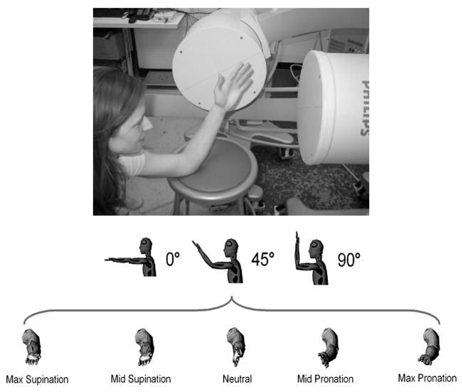
Methods: Changes in radioulnar kinematics during forearm supination-pronation and elbow flexion (0 degrees to 90 degrees ) were studied in 5 uninjured subjects using computed tomography, dual-orthogonal fluoroscopy, and 3-dimensional modeling. Analysis of variance and post-hoc testing was performed.
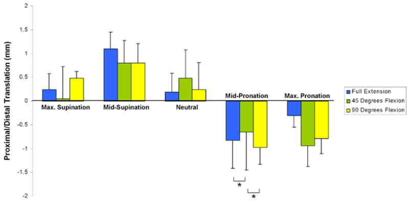
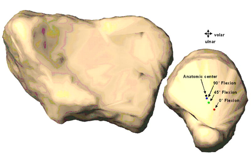
Results: Proximal translation of the radius was greatest with the elbow flexed to 90 degrees with the arm in midpronation. With the arm in midpronation, the translation of the radius was significantly greater at 0 degrees versus 45 degrees of elbow flexion (0.82 +/- 0.59 mm vs 0.65 +/- 0.80 mm, F: 4.49, post hoc: 0.055; p = .05) and significantly smaller at 45 degrees versus 90 degrees of elbow flexion (0.65 +/- 0.80 mm vs 0.97 +/- 0.35 mm, F: 4.49, post hoc: 0.048; p = .05). Proximal translation of the radius in midpronation was significantly greater than when the forearm was in a supinated position when the elbow was at 0 degrees or 90 degrees flexion (F: 14.90, post hoc: <0.01; p < .01, F: 19.11, post hoc: <0.01, p < .01). The arc of forearm rotation was significantly decreased at 0 degrees compared with 90 degrees of elbow flexion (129.3 degrees +/- 22.2 degrees vs 152.8 degrees +/- 14.4 degrees , F: 3.29, post hoc: 0.79; p = .09). The center of rotation shifted volarly and ulnarly with increasing elbow extension.
Conclusions: Elbow position affects the kinematics of the distal radioulnar joint. The kinematics of the distal radioulnar joint are primarily affected by forearm rotation and secondarily by elbow flexion. These findings have clinical relevance to our understanding of ulnar impaction, and how elbow position affects the proximal-distal translation of the radius. These findings have implications for the treatment of ulna impaction, radiographic evaluation of the distal ulna, and future biomechanical studies.
Figures
References
-
- Schuurman AH, Maas M, Dijkstra PF, Kauer JM. Assessment of ulnar variance: a radiological investigation in a Dutch population. Skeletal Radiol. 2001;30:633–8. - PubMed
-
- Yeh GL, Beredjiklian PK, Katz MA, Steinberg DR, Bozentka DJ. Effects of forearm rotation on the clinical evaluation of ulnar variance. J Hand Surg. 2001;26A:1042–1046. - PubMed
-
- Epner RA, Bowers WH, Guilford WB. Ulnar variance—the effect of wrist positioning and roentgen filming technique. J Hand Surg. 1982;7:298 –305. - PubMed
-
- Palmer AK, Werner FW. Biomechanics of the distal radioulnar joint. Clin Orthop. 1984;187:26 –35. - PubMed
-
- Tay SC, Berger RA, Tomita K, Tan ET, Amrami KK, An KN. In vivo three-dimensional displacement of the distal radioulnar joint during resisted forearm rotation. J Hand Surg. 2007;32A:450–8. - PubMed
Publication types
MeSH terms
Grants and funding
LinkOut - more resources
Full Text Sources
