. 2010 Aug;21(8):687-94.
doi: 10.1016/j.jnutbio.2009.04.002. Epub 2009 Jul 2.

Differential response to zinc-induced apoptosis in benign prostate hyperplasia and prostate cancer cells

Affiliations

Differential response to zinc-induced apoptosis in benign prostate hyperplasia and prostate cancer cells

Michelle Yan et al. J Nutr Biochem. 2010 Aug.

Abstract

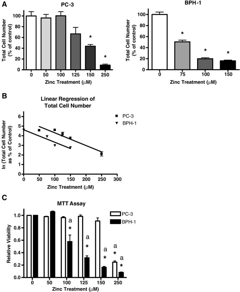
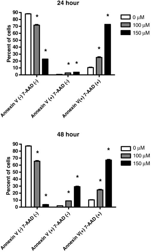
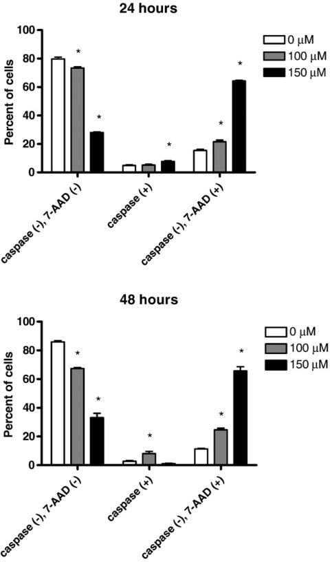
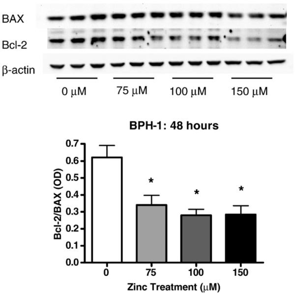
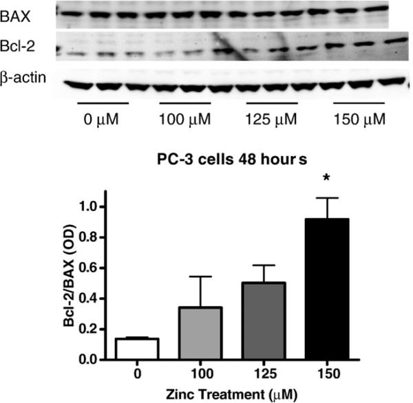
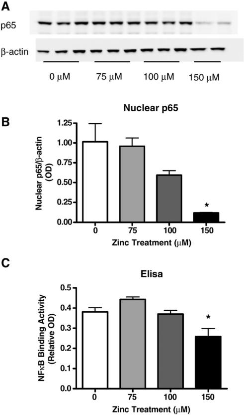
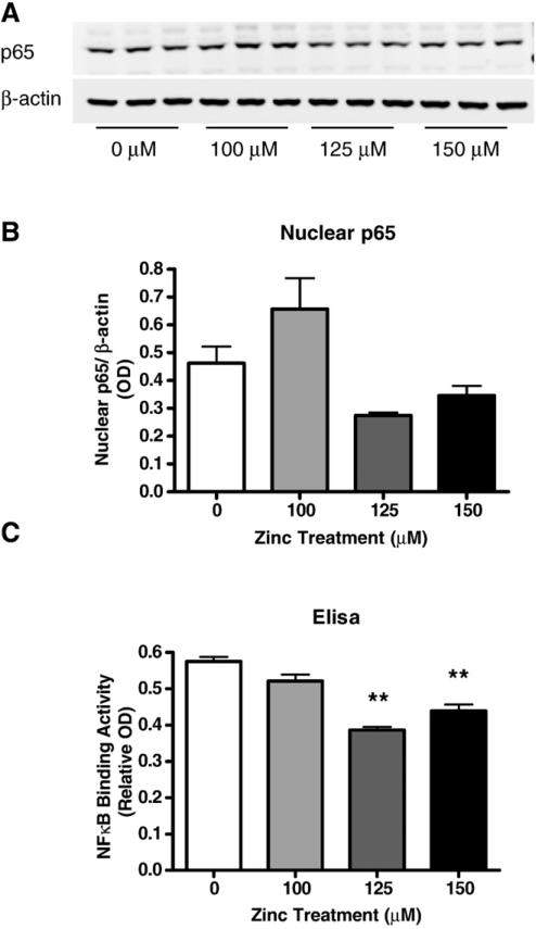
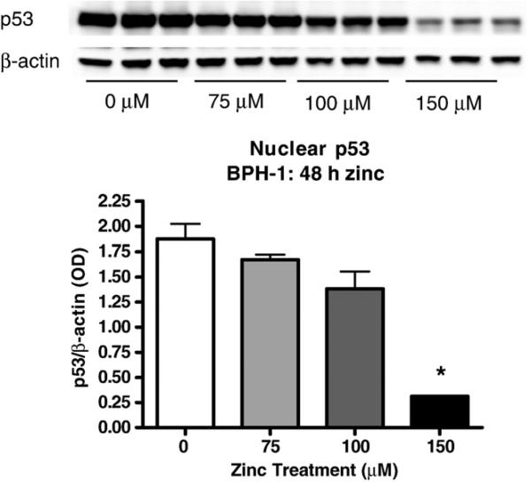
Zinc concentrations in the prostate are uniquely high but are dramatically decreased with prostate cancer. Studies have suggested that increasing zinc in the prostate may be a potential therapeutic strategy. The goal of this study was to evaluate the antiproliferative effects of zinc in prostate cancer cells (PC-3) and noncancerous benign prostate hyperplasia (BPH) cells (BPH-1) and to define possible mechanisms. PC-3 and BPH-1 cells were treated with zinc (0-250 microM) for 24 and 48 h, and cell growth and viability were examined. Apoptosis was assessed by phosphatidylserine externalization, caspase activation and protein expression of B-cell CLL/lymphoma 2 (Bcl-2)-associated X protein (BAX):Bcl-2. BPH-1 cells were more sensitive to the antiproliferative effects of zinc compared to PC-3. The response to zinc in PC-3 and BPH-1 cells differed as evidenced by opposing effects on Bcl-2:BAX expression. Additionally, different effects on the nuclear expression and activity of the p65 subunit of nuclear factor kappa B were observed in response to zinc between the two cell types. The differential response to zinc in PC-3 and BPH-1 cells suggests that zinc may serve an important role in regulating cell growth and apoptosis in prostate cancer and hyperplasia cells.


Figures

Fig. 1
Fig. 1
BPH-1 cells were more sensitive to the antiproliferative effects of zinc compared to PC-3. Cells were treated with zinc for 48 h; total cell numbers were determined using Beckman Coulter Z1 Coulter Particle Counter and plotted as percent of control±S.E.M. (A) Higher concentrations of zinc (150 μM) were required to significantly decrease total cell number in PC-3 compared to BPH-1 cells (75 μM). (B) Linear regression lines were determined for PC-3 and BPH-1 cells, and data is shown as percent of total cells as a function of zinc treatment. (C) PC-3 and BPH-1 cells were treated with zinc for 24 h, and viability was determined by MTT assay. Higher concentrations of zinc were required to induce a significant decrease in cell viability in PC-3 compared to BPH-1 cells. *P<.05 versus control treatment (no zinc) within a cell type. “A” indicates significant difference between PC-3 and BPH-1 cells within treatment.
Fig. 2
Fig. 2
Zinc increased annexin V staining, a marker of apoptosis, in BPH-1 cells. Cells were stained with annexin V and 7-AAD (an indicator of membrane integrity). Annexin V (-) 7-AAD (−) represents viable cells, annexin V (+) 7-AAD (−) represents early apoptotic cells and annexin V (+) 7-AAD (−) represents late apoptotic/necrotic cells. Both concentrations of zinc significantly decreased percentage of viable cells and increased apoptotic and necrotic cells. Statistical analysis was conducted using one-way ANOVA to compare zinc treatments with control (0 μM zinc) within each staining pattern [annexin V (−) 7-AAD (-), annexin V (+) 7-AAD (−) and annexin V (+) 7-AAD (+)] for each time point. Asterisk indicates P<.001. Triplicate samples were analyzed and results plotted as mean±S.E.M.
Fig. 3
Fig. 3
Zinc induced caspase activation, decreased cell viability and increased cell death in BPH-1 cells. Cells were analyzed for caspase activation using the Guava Multi Caspase Kit. Cells were exposed to the SR-VAD-FMK fluorochrome (which covalently binds to activated caspases) and the cell impermeant dye 7-AAD (an indicator of cell membrane integrity). Caspase (-) 7-AAD (−) represents viable cells; Caspase (+) represents early and late apoptotic cells; and caspase (−), 7-AAD (+) represents late apoptotic/necrotic cells. Cultures treated with zinc have reduced cell viability, increased caspase activation and an increased population of apoptotic and necrotic cells. Statistical analysis was conducted using one-way ANOVA, *P<.05 compared to 0 μM zinc within each treatment for each staining pattern for each time point. Triplicate samples were analyzed and results plotted as mean±S.E.M.
Fig. 4
Fig. 4
Zinc caused a significant decrease in Bcl-2:BAX ratio after 48 h in BPH-1 cells. BAX and Bcl-2 protein expression was determined by Western blotting. B-actin was used as the loading control. Triplicate samples were analyzed. Densitometry was conducted to quantify protein expression and plotted as OD (normalized to actin) versus zinc treatment as mean±S.E.M. Statistical analysis was conducted using oneway ANOVA, comparing each treatment to 0 μM zinc treatment. Triplicate samples were analyzed. BAX expression was significantly decreased at ≥100 μM zinc. Bcl-2 expression was significantly decreased at ≥75 μM zinc. The Bcl-2:BAX ratio was significantly decreased at ≥75 μM zinc. Asterisk indicates P<.05.
Fig. 5
Fig. 5
Zinc induced a significant increase in Bcl-2:BAX ratio after 48 h in PC-3. BAX and Bcl-2 protein expression was determined by Western blotting. B-actin was used as the loading control. Triplicate samples were analyzed. Densitometry was conducted to quantify protein expression and plotted as OD versus zinc treatment as mean±S.E. Statistical analysis was conducted using one-way ANOVA, comparing each treatment to 0 μM zinc treatment. Triplicate samples were analyzed. There was no significant change in BAX or Bcl-2 alone. There was a significant increase in Bcl-2:BAX expression at 150 μM zinc. Asterisk indicates P<.05.
Fig. 6
Fig. 6
Zinc decreased nuclear p65 expression and binding activity in BPH-1 cells. BPH-1 cells were treated with zinc for 48 h. Nuclear protein expression of p65 was determined by Western blotting (A), quantified by densitometry analysis and plotted as mean±S.E. (B). Beta-actin was used as the loading control; 150 μM zinc induced a significant decrease in p65 nuclear protein expression after 48 hours (P<.05). (C) Accordingly, 150 μM zinc also decreased p65 binding activity as determined by ELISA (P<.05).
Fig. 7
Fig. 7
Zinc did not change nuclear p65 expression in PC-3 cells. PC-3 were treated with zinc for 48 h. Nuclear protein expression of p65 was determined by Western blotting, quantified by densitometry analysis and plotted as mean±S.E. Beta-actin was used as the loading control. Zinc concentrations of ≥125 μM resulted in increased p65 binding activity as determined by ELISA (P<.01).
Fig. 8
Fig. 8
Zinc decreases nuclear p53 expression in BPH-1 cells. BPH-1 cells were treated with 0–150 mM zinc for 48 h and then harvested for analysis. Nuclear p53 expression was determined by Western blotting, quantified by densitometry analysis and plotted as mean±S.E. Triplicate samples were analyzed. One-way ANOVA was conducted with Tukey's multiple comparison testing. Asterisk indicates P<.001 compared to 0 μM zinc treatment.

References

    1. American Cancer Society . Cancer Facts & Figures 2008. American Cancer Society; Atlanta: 2008.
    1. Kirby RS. The natural history of benign prostatic hyperplasia: what have we learned in the last decade? Urology. 2000;56:3–6. - PubMed
    1. Clark LC, Combs GF, Jr, Turnbull BW, Slate EH, Chalker DK, Chow J, et al. Effects of selenium supplementation for cancer prevention in patients with carcinoma of the skin. A randomized controlled trial. Nutritional Prevention of Cancer Study Group. JAMA. 1996;276:1957–63. - PubMed
    1. Giovannucci E, Ascherio A, Rimm EB, Stampfer MJ, Colditz GA, Willett WC. Intake of carotenoids and retinol in relation to risk of prostate cancer. J Natl Cancer Inst. 1995;87:1767–76. - PubMed
    1. Giovannucci E, Rimm EB, Liu Y, Stampfer MJ, Willett WC. A prospective study of tomato products, lycopene, and prostate cancer risk. J Natl Cancer Inst. 2002;94:391–8. - PubMed

Publication types