Neuroprotective effects of the complement terminal pathway during demyelination: implications for oligodendrocyte survival
- PMID: 19577811
- PMCID: PMC2756021
- DOI: 10.1016/j.jneuroim.2009.06.006
Neuroprotective effects of the complement terminal pathway during demyelination: implications for oligodendrocyte survival
Abstract
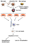
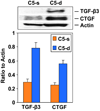
Multiple sclerosis (MS) is a chronic inflammatory demyelinating disease of the central nervous system that is mediated by activated lymphocytes, macrophages/microglia, and complement. In MS, the myelin-forming oligodendrocytes (OLGs) are the targets of the immune attack. Experimental evidence indicates that C5b-9 plays a role in demyelination during the acute phase of experimental allergic encephalomyelitis (EAE). Terminal complement C5b-9 complexes are capable of protecting OLGs from apoptosis. During chronic EAE complement C5 promotes axonal preservation, remyelination and provides protection from gliosis. These findings indicate that the activation of complement and C5b-9 assembly can also have protective roles during demyelination.
Figures


Similar articles
-
Complement activation in autoimmune demyelination: dual role in neuroinflammation and neuroprotection.J Neuroimmunol. 2006 Nov;180(1-2):9-16. doi: 10.1016/j.jneuroim.2006.07.009. Epub 2006 Aug 14. J Neuroimmunol. 2006. PMID: 16905199 Review.
-
C5b-9 complement complex in autoimmune demyelination and multiple sclerosis: dual role in neuroinflammation and neuroprotection.Ann Med. 2005;37(2):97-104. doi: 10.1080/07853890510007278. Ann Med. 2005. PMID: 16026117 Review.
-
Oligodendrocyte cell death in pathogenesis of multiple sclerosis: Protection of oligodendrocytes from apoptosis by complement.J Rehabil Res Dev. 2006 Jan-Feb;43(1):123-32. doi: 10.1682/jrrd.2004.08.0111. J Rehabil Res Dev. 2006. PMID: 16847778 Review.
-
Effects of complement C5 on apoptosis in experimental autoimmune encephalomyelitis.J Immunol. 2004 May 1;172(9):5702-6. doi: 10.4049/jimmunol.172.9.5702. J Immunol. 2004. PMID: 15100315
-
The response of NG2-expressing oligodendrocyte progenitors to demyelination in MOG-EAE and MS.J Neurocytol. 2002 Jul-Aug;31(6-7):523-36. doi: 10.1023/a:1025747832215. J Neurocytol. 2002. PMID: 14501221
Cited by
-
Microbial Neuraminidase Induces a Moderate and Transient Myelin Vacuolation Independent of Complement System Activation.Front Neurol. 2017 Mar 7;8:78. doi: 10.3389/fneur.2017.00078. eCollection 2017. Front Neurol. 2017. PMID: 28326060 Free PMC article.
-
Olfactory impairment in patients with primary Sjogren's syndrome and its correlation with organ involvement and immunological abnormalities.Arthritis Res Ther. 2021 Sep 29;23(1):250. doi: 10.1186/s13075-021-02624-6. Arthritis Res Ther. 2021. PMID: 34587995 Free PMC article.
-
Role of SIRT1 in autoimmune demyelination and neurodegeneration.Immunol Res. 2015 Mar;61(3):187-97. doi: 10.1007/s12026-014-8557-5. Immunol Res. 2015. PMID: 25281273 Review.
-
An ανβ3 integrin-binding peptide ameliorates symptoms of chronic progressive experimental autoimmune encephalomyelitis by alleviating neuroinflammatory responses in mice.J Neuroimmune Pharmacol. 2014 Jun;9(3):399-412. doi: 10.1007/s11481-014-9532-6. Epub 2014 Feb 28. J Neuroimmune Pharmacol. 2014. PMID: 24577603
-
The effect of smoking on the symptoms and progression of multiple sclerosis: a review.J Inflamm Res. 2010;3:115-26. doi: 10.2147/JIR.S12059. Epub 2010 Sep 1. J Inflamm Res. 2010. PMID: 22096361 Free PMC article.
References
-
- Akassoglou K, Bauer J, Kassiotis G, Pasparakis M, Lassmann H, Kollias G, Probert L. Oligodendrocyte apoptosis and primary demyelination induced by local TNF/p55TNF receptor signaling in the central nervous system of transgenic mice: models for multiple sclerosis with primary oligodendrogliopathy. Am J Pathol. 1998;153:801–813. - PMC - PubMed
-
- Andrews T, Zhang P, Bhat NR. TNFalpha potentiates IFNgamma-induced cell death in oligodendrocyte progenitors. J Neurosci Res. 1998;54:574–583. - PubMed
-
- Antel JP, McCrea E, Ladiwala U, Qin YF, Becher B. Non-MHC-restricted cell-mediated lysis of human oligodendrocytes in vitro: relation with CD56 expression. J Immunol. 1998;160:1606–1611. - PubMed
-
- Ashburner M, Ball CA, Blake JA, Botstein D, Butler H, Cherry JM, Davis AP, Dolinski K, Dwight SS, Eppig JT, Harris MA, Hill DP, Issel-Tarver L, Kasarskis A, Lewis S, Matese JC, Richardson JE, Ringwald M, Rubin GM, Sherlock G. Gene ontology: tool for the unification of biology The Gene Ontology Consortium. Nat Genet. 2000;25:25–29. - PMC - PubMed
Publication types
MeSH terms
Substances
Grants and funding
LinkOut - more resources
Full Text Sources
Medical
Miscellaneous

