

Proteasome inhibitors and cardiac cell growth
- PMID: 19578073
- PMCID: PMC2797448
- DOI: 10.1093/cvr/cvp226
Proteasome inhibitors and cardiac cell growth
Abstract
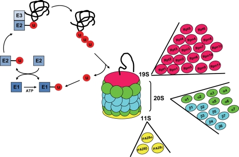
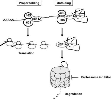
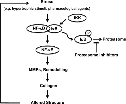
Activation of the ubiquitin-proteasome system has been described in different models of cardiac hypertrophy. Cardiac cell growth in response to pressure or volume overload, as well as physiological adaptive hypertrophy, is accompanied by an increase in protein ubiquitination, proteasome subunit expression, and proteasome activity. Importantly, an inhibition of proteasome activity prevents and reverses cardiac hypertrophy and remodelling in vivo. The focus of this review is to provide an update about the mechanisms by which proteasome inhibitors affect cardiac cell growth in adaptive and maladaptive models of cardiac hypertrophy. In the first part, we summarize how the proteasome affects both proteolysis and protein synthesis in a context of cardiac cell growth. In the second part, we show how proteasome inhibition can prevent and reverse cardiac hypertrophy and remodelling in response to different conditions of overload.
Figures
References
-
- Ciechanover A, Hod Y, Hershko A. A heat-stable polypeptide component of an ATP-dependent proteolytic system from reticulocytes. Biochem Biophys Res Commun. 1978;81:1100–1105. - PubMed
-
- Hershko A, Heller H, Elias S, Ciechanover A. Components of ubiquitin-protein ligase system. Resolution, affinity purification, and role in protein breakdown. J Biol Chem. 1983;258:8206–8214. - PubMed
-
- Meiners S, Ludwig A, Stangl V, Stangl K. Proteasome inhibitors: poisons and remedies. Med Res Rev. 2008;28:309–327. - PubMed
-
- Powell SR. The ubiquitin-proteasome system in cardiac physiology and pathology. Am J Physiol Heart Circ Physiol. 2006;291:H1–H19. - PubMed
