

Epileptogenesis in the immature brain: emerging mechanisms
- PMID: 19578345
- PMCID: PMC2822660
- DOI: 10.1038/nrneurol.2009.80
Epileptogenesis in the immature brain: emerging mechanisms
Abstract
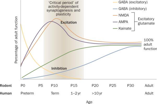
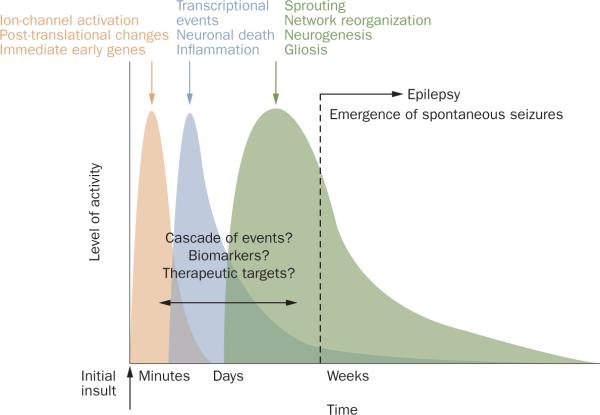
Epileptogenesis is defined as the process of developing epilepsy-a disorder characterized by recurrent seizures-following an initial insult. Seizure incidence during the human lifespan is at its highest in infancy and childhood. Animal models of epilepsy and human tissue studies suggest that epileptogenesis involves a cascade of molecular, cellular and neuronal network alterations. Within minutes to days following the initial insult, there are acute early changes in neuronal networks, which include rapid alterations to ion channel kinetics as a result of membrane depolarization, post-translational modifications to existing functional proteins, and activation of immediate early genes. Subacute changes occur over hours to weeks, and include transcriptional events, neuronal death and activation of inflammatory cascades. The chronic changes that follow over weeks to months include anatomical changes, such as neurogenesis, mossy fiber sprouting, network reorganization, and gliosis. These epileptogenic processes are developmentally regulated and might contribute to differences in epileptogenesis between adult and developing brains. Here we review the factors responsible for enhanced seizure susceptibility in the developing brain, and consider age-specific mechanisms of epileptogenesis. An understanding of these factors could yield potential therapeutic targets for the prevention of epileptogenesis and also provide biomarkers for identifying patients at risk of developing epilepsy or for monitoring disease progression.
Figures
References
-
- Hauser WA, Annegers JF, Kurland LT. Incidence of epilepsy and unprovoked seizures in Rochester, Minnesota: 1935–1984. Epilepsia. 1993;34:453–468. - PubMed
-
- Volpe JJ. Neurology of the Newborn. Saunders; Philadelphia: 2008. pp. 203–244.
-
- LaFrance WC, Jr, Kanner AM, Hermann B. Psychiatric comorbidities in epilepsy. Int. Rev. Neurobiol. 2008;83:347–383. - PubMed
-
- Temkin NR. Antiepileptogenesis and seizure prevention trials with antiepileptic drugs: meta-analysis of controlled trials. Epilepsia. 2001;42:515–524. - PubMed
Publication types
MeSH terms
Grants and funding
LinkOut - more resources
Full Text Sources
Other Literature Sources
Medical
