

doi: 10.1038/nmeth.1347.
Epub 2009 Jul 5.
Mass spectrometry of membrane transporters reveals subunit stoichiometry and interactions
Affiliations
- PMID: 19578383
- PMCID: PMC4066579
- DOI: 10.1038/nmeth.1347
Item in Clipboard
Mass spectrometry of membrane transporters reveals subunit stoichiometry and interactions
Nat Methods.
2009 Aug.
Abstract
We describe a general mass spectrometry approach to determine subunit stoichiometry and lipid binding in intact membrane protein complexes. By exploring conditions for preserving interactions during transmission into the gas phase and for optimally stripping away detergent, by subjecting the complex to multiple collisions, we released the intact complex largely devoid of detergent. This enabled us to characterize both subunit stoichiometry and lipid binding in 4 membrane protein complexes.
Figures
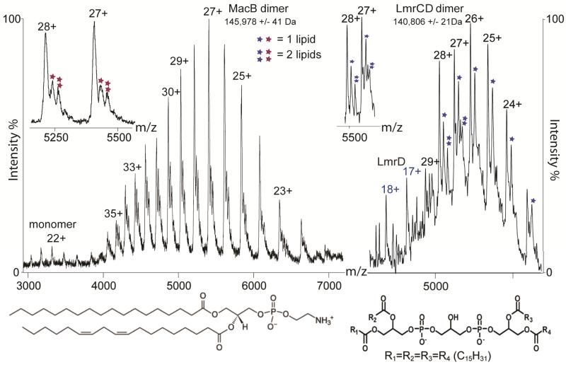
Mass spectra of MacB (left) and LmrCD (right) showing almost exclusively homodimer and heterodimer formation, respectively. Insets, expansion across charge states showing that phosphatidylenthanolamine and cardiolipin bind to the MacB and LmrCD dimers, respectively. Structures of phosphatidylenthanolamine (left) and cardiolipin (right) are shown at the bottom.
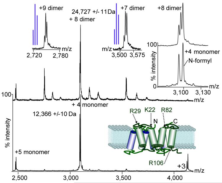
Mass spectra of EmrE-EFE(1–113) under conditions where interactions are lost (bottom spectrum) and preserved (top spectrum). Schematic representation of the EmrE based on a structure from the Protein Data Bank (PDB) (3B5D) shows the location of basic residues. Insets, expansion of the monomeric and dimeric charge states reveals modified (N-terminally formylated) and free protein. Blue lines indicate schematically the peaks intensities calculated for a statistical incorporation of subunits into the dimer.
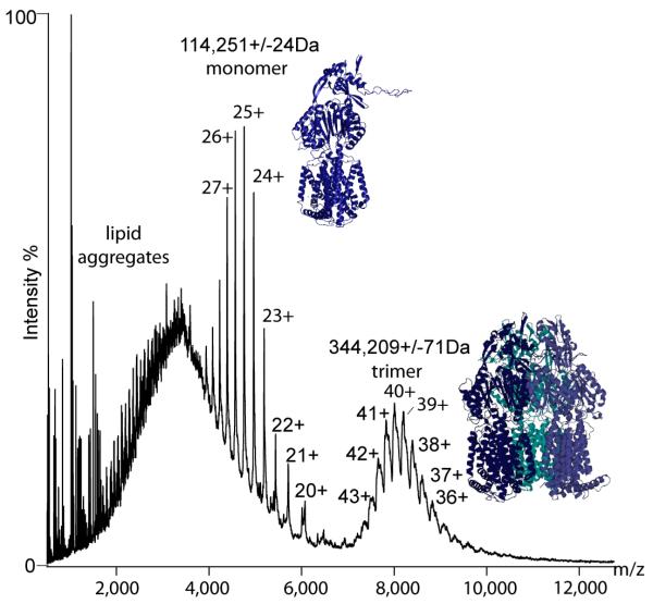
Mass spectrum of MexB, revealing only one oligomeric form, the trimer (right). Also present in the spectra are lipid aggregates and a well resolved monomeric form (left). Atomic models of the monomer were generated using 3D-jigsaw via homology modeling from the X-ray structure of AcrB. The model of trimeric MexB was generated using the program Modeller.
References
-
- Schagger H, von Jagow G. Anal. Biochem. 1991;199:223–231. - PubMed
-
- Burgess NK, Stanley AM, Fleming KG. Methods Cell Biol. 2008;84:181–211. - PubMed
-
- Putnam CD, Hammel M, Hura GL, Tainer JA. Q. Rev. Biophys. 2007;40:191–285. - PubMed
-
- Barrera NP, Di Bartolo N, Booth PJ, Robinson CV. Science. 2008;321:243–246. - PubMed
-
- Locher KP, Lee AT, Rees DC. Science. 2002;296:1091–1098. - PubMed
Publication types
MeSH terms
Substances
Grants and funding
LinkOut - more resources
Full Text Sources
Other Literature Sources
