

Endobronchial ultrasound application for diagnosis of tracheobronchial tree invasion by esophageal cancer
- PMID: 19578652
- PMCID: PMC2705145
- DOI: 10.1590/s1807-59322009000600003
Endobronchial ultrasound application for diagnosis of tracheobronchial tree invasion by esophageal cancer
Abstract
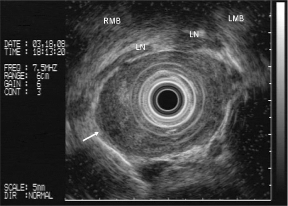
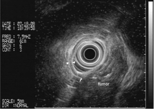
Introduction: Esophageal cancer staging has been performed through bronchoscopy, computerized tomography (CT), positron emission tomography (PET), and endoscopic ultrasound (EUS). Whereas CT and PET scan provide assessments of distant metastasis, bronchoscopy importantly diagnoses tracheobronchial involvement, complementing chest CT findings. EUS is the most accurate examination for T and N staging but is technically limited when tumoral stenoses cannot be traversed. Endobronchial ultrasound (EBUS) appears to present greater accuracy than EUS, CT, and bronchoscopy for assessing tracheobronchial wall involvement. EBUS has been recently associated with EUS for esophageal cancer staging in our unit.
Objective: To compare EBUS findings in esophageal cancer patients without evident signs of tracheobronchial invasion on conventional bronchoscopy with EUS and CT.
Methods: Fourteen patients with esophageal cancer underwent CT, conventional bronchoscopy, EUS, and EBUS for preoperative staging. All patients underwent EBUS and EUS with an Olympus(R) MH-908 echoendoscope at 7.5 MHz. Seven patients were eligible for the study according to the inclusion criteria.
Results: The echoendoscope could not traverse tumoral esophageal stenosis to perform EUS in two patients, and invasion was effectively diagnosed by EBUS. In 4 (57%) of 7 patients EBUS revealed additional information to staging. In the remaining 3 cases the invasion findings were the same under both EUS and EBUS.
Conclusion: EBUS showed signs of tracheobronchial invasion not observed by conventional bronchoscopy, adding information to staging in most of the cases when compared with CT and EUS.
Keywords: Endobronchial ultrasound; Endoscopic ultrasound; Esophageal neoplasia; Staging.
Figures
References
-
- Parada A.Tumores do EsôfagoIn: SOBED ed. Endoscopia Digestiva Rio de Janeiro, RJ: MEDSI Editora Médica e Científica Ltda; 43–56.
-
- Zilberstein B, Pinotti HW, Cecconello I, Ibrahim RE. Câncer do Esôfago. In: Pinotti HW, editor. Tratado de Clínica Cirúrgica do Aparelho Digestivo. 1 ed. São Paulo, SP: Atheneu; pp. 415–26.
-
- Roth JA, Lichter AS, Putnam JB. Cancer of the esophagus. In: DeVita VT, Hellman S, Rosenberg SA, editors. Cancer: Principles & Practice on Oncology. 4th edition ed. Philadelphia: Lippincott Williams & Wilkins; 1997. pp. 776–817.
-
- Bains MS, Shields TW. Squamous cell carcinoma of the esophagus. In: Shields TW, LoCicero J, Ponn RB, Rusch VW, editors. General thoracic surgery. 4th ed. Baltimore: Williams & Wilkins; 1994. pp. 1633–58.
-
- Räsänen JV, Sihvo EI, Knuuti MJ, Minn HR, Luostarinen ME, Laippala P, et al. Prospective analysis of accuracy of positron emission tomography, computed tomography, and endoscopic ultrasonography in staging of adenocarcinoma of the esophagus and the esophagogastric junction. Ann Surg Oncol. 2003;10:954–60. - PubMed
Publication types
MeSH terms
LinkOut - more resources
Full Text Sources
Medical
