. 2009 Aug;35(2):337-45.

MUC1 is a downstream target of STAT3 and regulates lung cancer cell survival and invasion

Affiliations

MUC1 is a downstream target of STAT3 and regulates lung cancer cell survival and invasion

Jingchun Gao et al. Int J Oncol. 2009 Aug.

Abstract

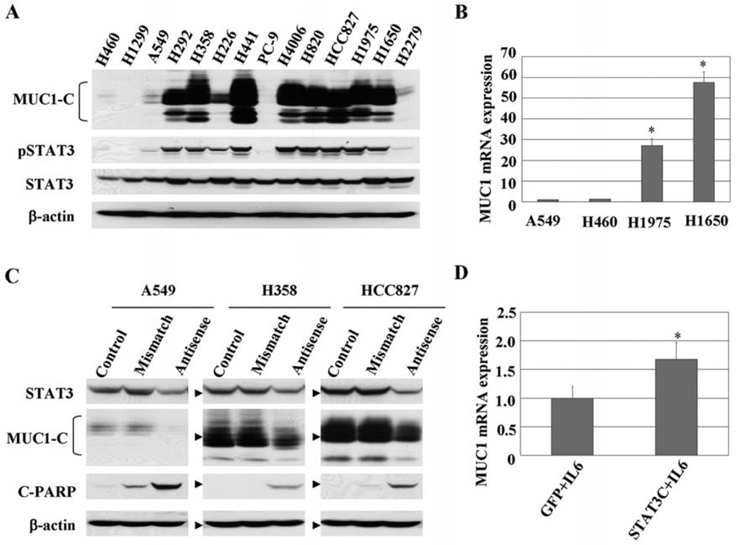
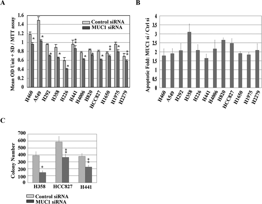
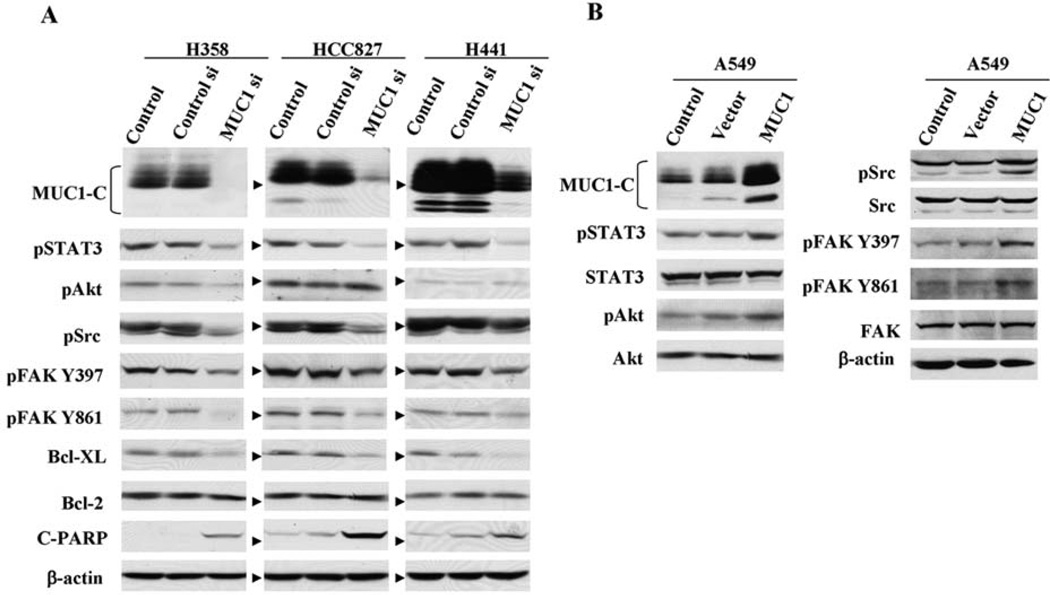
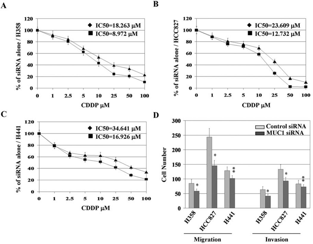
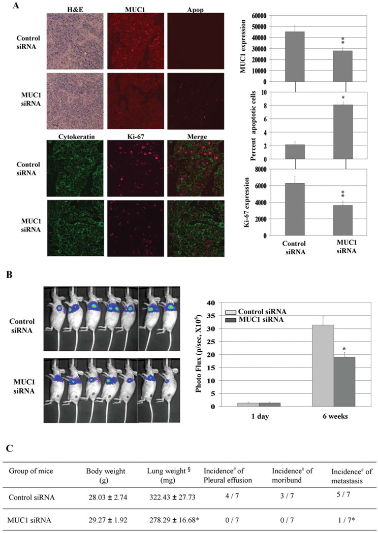
Signal transducer and activator of transcription 3 (STAT3) is aberrantly activated in human cancer including lung cancer and has been implicated in transformation, tumorigenicity, and metastasis. One putative downstream gene regulated by Stat3 is MUC1 which also has important roles in tumorigenesis. We determined if Stat3 regulates MUC1 in lung cancer cell lines and what function MUC1 plays in lung cancer cell biology. We examined MUC1 expression in non-small cell lung cancer (NSCLC) cell lines and found high levels of MUC1 protein expression associated with higher levels of tyrosine phosphorylated STAT3. STAT3 knockdown downregulated MUC1 expression whereas constitutive STAT3 expression increased MUC1 expression at mRNA and protein levels. MUC1 knockdown induced cellular apoptosis concomitant with reduced Bcl-XL and sensitized cells to cisplatin treatment. MUC1 knockdown inhibited tumor growth and metastasis in an orthotopic mouse model of lung cancer by activating apoptosis and inhibiting cell proliferation in vivo. These results demonstrate that constitutively activated STAT3 regulates expression of MUC1, which mediates lung cancer cell survival and metastasis in vitro and in vivo. MUC1 appears to be a cooperating oncoprotein with multiple oncogenic tyrosine kinase pathways and could be an effective target for the treatment of lung cancer.


Figures

Figure 1
Figure 1
MUC1 is widely overexpressed in NSCLC cells, correlating with STAT3 activation. (A) Protein expression of MUC1-C and total and Tyr705 phosphorylated STAT3 levels were determined in 14 human NSCLC cell lines by Western blot analyses. Equal loading and transfer were shown by repeat probing with β-actin. (B) mRNA levels of MUC1 in a subset of cells were evaluated with real-time RT-PCR. MUC1 mRNA expression levels in each cell line were normalized to GAPDH mRNA, with expression in A549 cells set to an arbitrary value of 1. *P<0.01 compared with A549 or H460 cells. (C) Cells were treated with 700 nM antisense STAT3 oligonucleotides for ~24–48 h, and the effects on MUC1 expression and PARP cleavage were analyzed by Western blot analysis. Equal loading and transfer were shown by repeat probing with β-actin. (D) A549 cells were infected with either control adenovirus (Ad-GFP) or Ad- Stat3C for 13 h along with low levels of IL-6 for an additional 5 h to enhance Stat3C tyrosine phosphorylation. mRNA levels of MUC1 expression were determined by real-time RT-PCR assay and normalized to GAPDH mRNA, with expression in Ad-GFP cells set to an arbitrary value of 1. *P<0.01, **P<0.05 compared with Ad-GFP cells.
Figure 2
Figure 2
MUC1 functions to drive cell survival and colony formation. (A) Cells were exposed to siRNA for 120 h, and proliferation rates were measured by MTT assay. *P<0.01, **P<0.05 compared with the control. (B) Cells were exposed to siRNA for 72 h, and cell apoptosis evidenced by positive cells of active caspase-3 in the cells was assessed by flow cytometry analysis. Relative units of apoptosis for the control were set to 1; data for the MUC1 siRNA are shown as fold change relative to control siRNA. (C) Cells treated with siRNA were grown in RPMI-1640 medium supplied with 10% FBS, and colonies were counted after 14 days. *P<0.01, **P<0.05 compared with the control.
Figure 3
Figure 3
Effects of MUC1 knockdown or overexpression on multiple cell signals in NSCLC cells. (A) H358, HCC827, and H441 cells were exposed to siRNA for 72 h, and (B) A549 cells were transfected with MUC1-C plasmid for 48 h followed by measurement of phosphorylated tyrosine STAT3, Akt, Src, FAK, and apoptotic-related proteins by Western blot analysis. Equal loading and transfer were shown by repeat probing with β-actin.
Figure 4
Figure 4
MUC1 knockdown sensitizes NSCLC cells to cisplatin treatment and inhibits metastasis potential. H358 (A), HCC827 (B), and H441 (C) cells were treated with 20 nM siRNA for 72 h, collected and plated in 96-well plates at a density of 10,000 cells/well, and allowed to grow for ~18–24 h before a 48-h exposure to cisplatin in dose gradient. Percentage of cell viability was qualified by MTT assay. IC50 of cisplatin was calculated using Matlab software. ♦, control siRNA + CDDP; ■, MUC1 siRNA + CDDP. (D) MUC1 overexpressing cell lines (H358, HCC827, and H441) were transfected with 20 nM siRNA for 72 h and then assessed for migration and invasion as described in Materials and methods. *P<0.01, **P<0.05 compared with the control.
Figure 5
Figure 5
MUC1 knockdown induces apoptosis in vivo and inhibits orthotopic tumor growth in CD-1 nude mice. (A) Lung tissue specimens subjected to H&E staining and immunohistochemical analysis of MUC1 and Ki-67 expression and apoptosis (Apop). *P<0.01, **P<0.05 compared with the control. (B) Results of orthotopic tumor growth evaluation using Xenogen 200 imaging system. *P<0.01 compared with the control. (C) Seven mice for each group were sacrificed 6 weeks after tumor injection when control mice became moribund. §, weight designates lung and tumor weight; #, number of positive mice/number of treated mice; *P<0.05 compared with the control siRNA group.

References

    1. Bowman T, Garcia R, Turkson J, Jove R. STATs in oncogenesis. Oncogene. 2000;19:2474–2488. - PubMed
    1. Frank DA. STAT signaling in the pathogenesis and treatment of cancer. Mol Med. 1999;5:432–456. - PMC - PubMed
    1. Haura EB, Zheng Z, Song L, Cantor A, Bepler G. Activated epidermal growth factor receptor-Stat-3 signaling promotes tumor survival in vivo in non-small cell lung cancer. Clin Cancer Res. 2005;11:8288–8894. - PubMed
    1. Alvarez JV, Greulich H, Sellers WR, Meyerson M, Frank DA. Signal transducer and activator of transcription 3 is required for the oncogenic effects of non-small-cell lung mutations of the epidermal growth factor receptor. Cancer Res. 2006;66:3162–3168. - PubMed
    1. Yu H, Jove R. The STATs of cancer - new molecular targets come of age. Nat Rev. 2004;4:97–105. - PubMed

Publication types

MeSH terms