. 2010 Mar;15(2):165-81.
doi: 10.1007/s12192-009-0130-0. Epub 2009 Jul 5.

Thiolutin inhibits endothelial cell adhesion by perturbing Hsp27 interactions with components of the actin and intermediate filament cytoskeleton

Affiliations

Thiolutin inhibits endothelial cell adhesion by perturbing Hsp27 interactions with components of the actin and intermediate filament cytoskeleton

Yifeng Jia et al. Cell Stress Chaperones. 2010 Mar.

Abstract

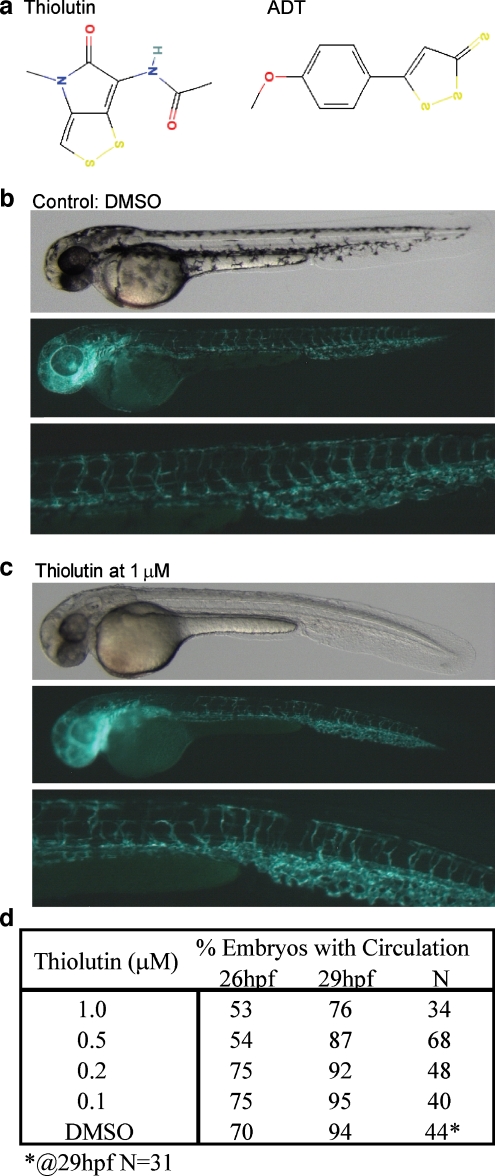
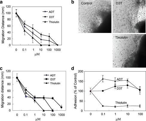
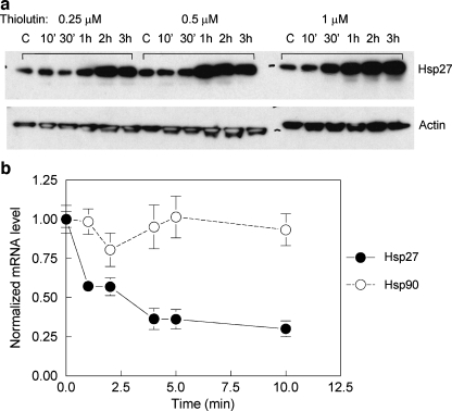
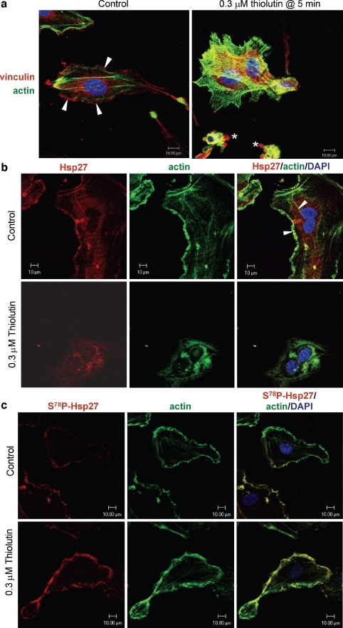
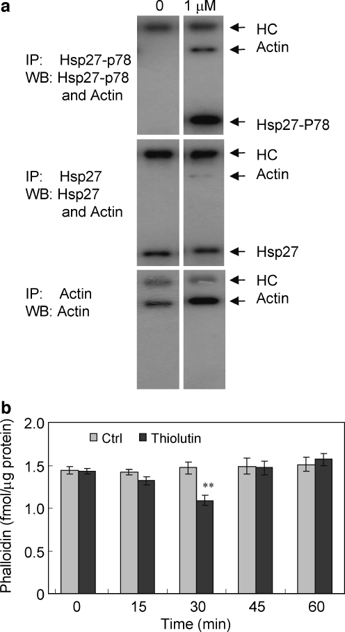
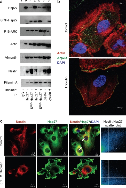
Thiolutin is a dithiole synthesized by Streptomyces sp. that inhibits endothelial cell adhesion and tumor growth. We show here that thiolutin potently inhibits developmental angiogenesis in zebrafish and vascular outgrowth from tissue explants in 3D cultures. Thiolutin is a potent and selective inhibitor of endothelial cell adhesion accompanied by rapid induction of HSPB1 (Hsp27) phosphorylation. The inhibitory effects of thiolutin on endothelial cell adhesion are transient, potentially due to a compensatory increase in Hsp27 protein levels. Accordingly, heat shock induction of Hsp27 limits the anti-adhesive activity of thiolutin. Thiolutin treatment results in loss of actin stress fibers, increased cortical actin as cells retract, and decreased cellular F-actin. Mass spectrometric analysis of Hsp27 binding partners following immunoaffinity purification identified several regulatory components of the actin cytoskeleton that associate with Hsp27 in a thiolutin-sensitive manner including several components of the Arp2/3 complex. Among these, ArpC1a is a direct binding partner of Hsp27. Thiolutin treatment induces peripheral localization of phosphorylated Hsp27 and Arp2/3. Hsp27 also associates with the intermediate filament components vimentin and nestin. Thiolutin treatment specifically ablates Hsp27 interaction with nestin and collapses nestin filaments. These results provide new mechanistic insights into regulation of cell adhesion and cytoskeletal dynamics by Hsp27.


Figures

Fig. 1
Fig. 1
Thiolutin inhibits zebrafish embryonic development and angiogenesis. a Structures of thiolutin and ADT. Both have αβ-unsaturated dithiole moieties. bc Lateral whole mount views of zebrafish embryos treated with DMSO (b) or thiolutin (c). Each panel contains three pictures. The top picture in each panel is a phase contrast image, the middle picture is a fluorescent (GFP) image of the same embryo, and the bottom picture is the close up view of the trunk region of the treated embryo. Left is head and right is tail region. d At lower concentrations of thiolutin (less than 1 μM), 50% of embryos showed circulatory defects at 26 hpf when compared to 25% of age-matched DMSO treated embryos, but fewer embryos showed such defects at 29 hpf
Fig. 2
Fig. 2
Thiolutin blocks both wound-driven and tumor-driven vascular outgrowths. a, b C57BL6 murine muscle biopsies were explanted in three-dimensional collagen matrix and incubated in the presence of the indicated treatments and cell migration measured at day 7. c Biopsies from HT29 adenocarcinoma xenografts harvested from Cr:(NCr)-athymic nu fBR mice were similarly treated. Explants were imaged using a 20× objective. Results represent the mean±SD of three separate experiments. d HUVEC (1 × 104 cell/well) were plated in 96-well culture dishes pre-coated with type I collagen (5 μg/ml) in the presence of the indicated treatment agents. Following incubation for 1 h at 37°C plates were washed and the attached cells were fixed, developed, and read at 570 nm. Results are expressed as percentage of control and represent the mean ± SD of at least three separate experiments
Fig. 3
Fig. 3
Thiolutin disrupts cell attachment and focal adhesions. HUVEC were cultured in 24-well plates in EGM-2%FBS media to 90% confluence and then incubated overnight in EBM-1%FBS. a Cells were treated with thiolutin at 0.5 μM and digital images were acquired every 15 min. b Cells were treated with thiolutin at 0.5 μM for 1 or 12 h (top panel), and then cells were collected and placed into fresh media to recover for 24 h (bottom panel). c HUVEC were treated with thiolutin at the indicated concentrations 23 h following plating, and adhesion was assessed continuously by impedance measurement using an ACEA RT-CES and presented in cell index units. d Heat-shocked HUVEC were allowed to recover for 24 h and then treated with thiolutin. Adhesion was assessed over time by changes in impedance as in c. The experiments were repeated at least three times with duplicates. Identical results were observed for each experiment
Fig. 4
Fig. 4
Thiolutin treatment increases Hsp27 protein levels but decreases Hsp27 mRNA. HUVEC were cultured as described in the “Materials and methods” section and treated with thiolutin at 0.25, 0.5, and 1 μM for the timed indicated. a The expression of Hsp27 was examined by blotting using an anti-Hsp27 antibody. The membranes were reprobed with an anti-actin antibody for loading control. b Real-time RT-PCR was performed as described in the “Materials and methods” section. Changes in the levels of Hsp27 and Hsp90 mRNA were determined at the indicated times following thiolutin treatment by the normalizing to HPRT mRNA levels in each sample and presented as mean±SD of triplicates
Fig. 5
Fig. 5
Thiolutin disrupts focal adhesions and induces association of phosphorylated Hsp27 with cortical actin. Confocal images were obtained for untreated HUVEC and HUVEC treated with 300 nM thiolutin for 5 min. a Vinculin (red) and actin (green) and nuclei (blue) staining in untreated HUVEC (left) and in HUVEC treated with thiolutin (right). b Hsp27 (red) and actin (green) were imaged in untreated (upper) and treated HUVEC (lower panels). c Hsp27-S78P (red) and actin (green) were imaged in untreated HUVEC (upper panels) and in HUVEC treated with thiolutin for 5 min (lower panels). Scale bars = 10 μm
Fig. 6
Fig. 6
Thiolutin disrupts actin stress fibers and affects actin polymerization. a Lysates of HUVEC treated with 1 μM thiolutin for 60 min were prepared and equally divided for immunoprecipitation. Anti- Hsp27, Hsp27-S78P, and actin antibodies were used at a 1:5,000 dilution and incubated for 1 h at 4°C. Protein-G was then added at a 1:500 dilution and incubated overnight at 4°C. After three washes by a quick spin, the resulting immune complexes were dissolved into 1× SDS sample buffer, and Western blotting (WB) was performed using the indicated antibodies. HC Ig heavy chain. b HUVEC cells were treated with 0.25 μM thiolutin for the time indicated. After centrifugation, the cells were fixed with 4% paraformaldehyde and incubated with phalloidin at saturated concentration for binding to F-actin. The bound phalloidin was extracted using methanol and quantified using a fluorescence plate reader. The experiments were performed four times with triplicates. **p < 0.05
Fig. 7
Fig. 7
Thiolutin alters Hsp27 binding to actin cytoskeleton and intermediate filament regulatory components. a Control HUVEC (lanes 1, 2, 3, and 6) or HUVEC treated with 0.5 μM thiolutin for 60 min (lanes 4, 5, and 7) were lysed, and supernatants following centrifugation were immunoprecipitated using control IgG (lane 1), anti-Hsp27 (lanes 2 and 4), and Hsp27-S78P antibodies (lanes 3 and 5). Associated levels of the indicated Hsp27-binding proteins in the immunoprecipitates (lanes 25) or whole cell lysates (lanes 6 and 7) were determined by Western blotting using the indicated antibodies. b Actin (red) and Arp2/3 component p34 (green) were imaged by confocal microscopy in untreated HUVEC (top) and in HUVEC treated with 200 nM thiolutin for 5 min (bottom). Inset shows a higher magnification of the retracting cell edge. c Nestin (red) and Hsp27 (green) were imaged in untreated HUVEC (upper panels) and HUVEC treated using 100 nM thiolutin for 5 min (lower panels). Representative images are presented. Scale bar = 10 μm. Right panels show plots of pixel intensities for the red and green channels that were used to calculate colocalization coefficients

References

    1. Adams CC, Gross DS. The yeast heat shock response is induced by conversion of cells to spheroplasts and by potent transcriptional inhibitors. J Bacteriol. 1991;173:7429–7435. - PMC - PubMed
    1. Arrigo AP, Virot S, Chaufour S, Firdaus W, Kretz-Remy C, Diaz-Latoud C. Hsp27 consolidates intracellular redox homeostasis by upholding glutathione in its reduced form and by decreasing iron intracellular levels. Antioxid Redox Signal. 2005;7:414–422. doi: 10.1089/ars.2005.7.414. - DOI - PubMed
    1. Balasubramanian MK, Feoktistova A, McCollum D, Gould KL. Fission yeast Sop2p: a novel and evolutionarily conserved protein that interacts with Arp3p and modulates profilin function. Embo J. 1996;15:6426–6437. - PMC - PubMed
    1. Berrou E, Bryckaert M. Recruitment of protein phosphatase 2A to dorsal ruffles by platelet-derived growth factor in smooth muscle cells: Dephosphorylation of Hsp27. Exp Cell Res. 2008;315:836–848. doi: 10.1016/j.yexcr.2008.12.002. - DOI - PubMed
    1. Bitar KN. HSP27 phosphorylation and interaction with actin-myosin in smooth muscle contraction. Am J Physiol Gastrointest Liver Physiol. 2002;282:G894–903. - PubMed

Publication types

MeSH terms

LinkOut - more resources