

Expression of activated checkpoint kinase 2 and histone 2AX in exfoliative oral cells after exposure to ionizing radiation
- PMID: 19580484
- PMCID: PMC3575577
- DOI: 10.1667/RR1560.1
Expression of activated checkpoint kinase 2 and histone 2AX in exfoliative oral cells after exposure to ionizing radiation
Abstract
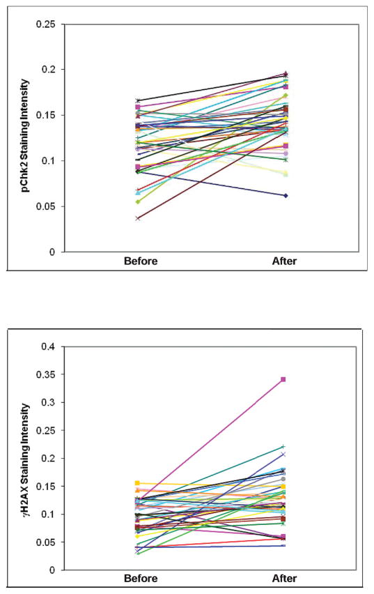
Gamma-H2AX (activated histone 2AX) and pChk2 (activated checkpoint kinase 2), which are DNA damage response molecules, are produced in irradiated cells and may be signature molecules of radiation exposure. We investigated their use as potential biomarkers to identify individuals exposed to ionizing radiation. We collected exfoliated oral epithelial cell samples from 100 healthy individuals undergoing routine dental radiographic examination (2.34 cGy) both before and after the radiograph using a non-invasive technique. The expression levels of pChk2 and gamma-H2AX in oral cells were assessed by immunohistochemical assay. Both biomarkers showed statistically significant increases in levels of expression after the radiation exposure (P < 0.001). This suggests that pChk2 and gamma-H2AX may serve as sensitive indicators of low-dose radiation exposure.
Figures


References
-
- Marchetti R, Coleman MA, Jones IM, Wyrobek AJ. Candidate protein biodosimeters of human exposure to ionizing radiation. Int J Radiat Biol. 2006;82:605–639. - PubMed
-
- Smilenov LB, Brenner DJ, Hall EJ. Modest increased sensitivity to radiation oncogenesis in ATM heterozygous versus wild-type mammalian cells. Cancer Res. 2001;61:5710–5713. - PubMed
-
- Bartkova J, Horejsi Z, Koed K, Kramer A, Tort F, Zieger K, Guldberg P, Sehested M, Nesland JM, Bartek J. DNA damage response as a candidate anti-cancer barrier in early human tumorigenesis. Nature. 2005;434:864–870. - PubMed
-
- Yarborough A, Zhang YJ, Hsu TM, Santella RM. Immunoperoxidase detection of 8-hydroxydeoxyguanosine in aflatoxin B1-treated rat liver and human oral mucosal cells. Cancer Res. 1996;56:683–688. - PubMed
Publication types
MeSH terms
Substances
Grants and funding
LinkOut - more resources
Full Text Sources
