. 2009 Nov;168(2):352-6.
doi: 10.1016/j.jsb.2009.07.001. Epub 2009 Jul 4.

Crystal structure of the in vivo-assembled Bacillus subtilis Spx/RNA polymerase alpha subunit C-terminal domain complex

Affiliations

Crystal structure of the in vivo-assembled Bacillus subtilis Spx/RNA polymerase alpha subunit C-terminal domain complex

Valerie Lamour et al. J Struct Biol. 2009 Nov.

Abstract

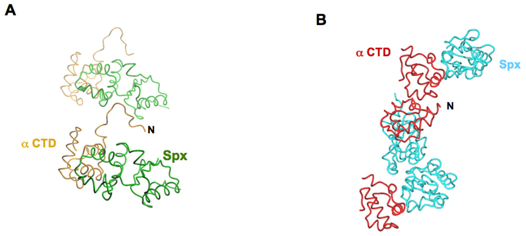
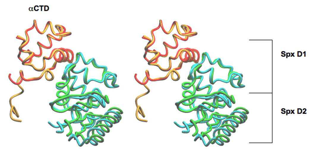
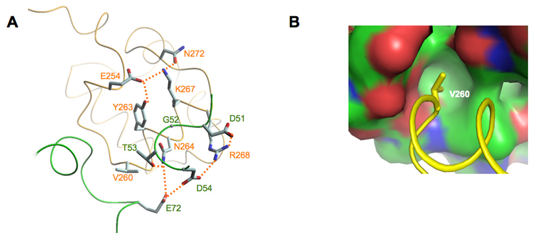
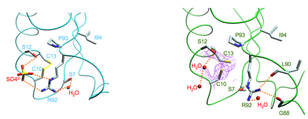
The Bacillus subtilis Spx protein is a global transcription factor that interacts with the C-terminal domain of the RNA polymerase alpha subunit (alphaCTD) and regulates transcription of genes involved in thiol-oxidative stress, sporulation, competence, and organosulfur metabolism. Here we determined the X-ray crystal structure of the Spx/alphaCTD complex from an entirely new crystal form than previously reported [Newberry, K.J., Nakano, S., Zuber, P., Brennan, R.G., 2005. Crystal structure of the Bacillus subtilis anti-alpha, global transcriptional regulator, Spx, in complex with the alpha C-terminal domain of RNA polymerase. Proc. Natl. Acad. Sci. USA 102, 15839-15844]. Comparison of the previously reported sulfate-bound complex and our sulfate-free complex reveals subtle conformational changes that may be important for the role of Spx in regulating organosulfur metabolism.


Figures

Figure 1
Figure 1
Crystal packing interactions. A. Crystal packing of the in vivo-assembled Spx/αCTD complex: the N-terminus of αCTD (labeled N) docks into a crevice formed between the two domains of a symmetry related Spx molecule. B. Crystal packing of the in vitro-assembled Spx/αCTD complex: crystal packing is maintained by both αCTD/αCTD interactions and Spx/Spx interactions. The in vivo-assembled complex αCTD is colored orange and its cognate Spx molecule is colored green. The in vitro-assembled complex αCTD is colored red and its cognate Spx molecule is colored cyan.
Figure 2
Figure 2
Stereoview of the superimposition of the in vivo- and in vitro-assembled complexes, crystallized in space groups P212121 and R3, respectively. The superimposition using LSQ was over the αCTD domain only (lsq_exp command, http://xray.bmc.uu.se/~alwyn/Essential_O/lsq_frameset.html). The N-terminus of αCTD in the in vivo-assembled complex is nine residues longer than in the in vitro-assembled complex. The αCTD molecules superimpose with a RMSD of 0.504 Å over 62 α carbon positions and the Spx molecules superimpose with a RMSD of 0.732 Å over 118 α carbon positions. In the in vivo-assembled complex αCTD is colored orange and Spx is colored green; in the in vitro-assembled complex αCTD is colored red and the Spx is colored cyan.
Figure 3
Figure 3
Molecular details of the Spx/αCTD interface. A. In both the in vivo- and the in vitro-assembled structures, αCTD residue Asn264 and Arg268 interact with Spx residues Asp51, Thr53 and Asp54. The residues that contribute to the overall Spx/αCTD interface are highlighted (hydrogen bonds are shown as dashed lines). B. αCTD residue Val260 projects into a hydrophobic pocket in Spx (the surface is colored in green for carbon atoms, red for oxygens and blue for nitrogens).
Figure 4
Figure 4
The “sulfate-free” conformation of Spx. A. In the structure of the in vitro-assembled complex (Newberry et al., 2005), a sulfate ion binds to Spx residues Ser12, which is adjacent to the disulfide bond formed between residues Cys10 and Cys13, and Arg92, which is part of the conserved Arg-Pro-Ile motif (Arg92, Pro93, Ile94). B. In the structure of the in vivo-assembled complex, no sulfate ion is present. The amine group of Arg92 is oriented “away” from the disulfide bond and forms an interaction with Ser7 and the carbonyl groups of residues Gly88 and Leu90. It is clear that residues Cys10 and Cys13 are oxidized, as revealed by the 2fo-fc electronic density (colored purple and contoured at 1.3σ).

References

    1. Benoff B, Yang H, Lawson CL, Parkinson G, Liu J, Blatter E, Ebright YW, Berman HM, Ebright RH. Structural basis of transcription activation: The CAP-αCTD-DNA complex. Science. 2002;297:1562–1566. - PubMed
    1. Brünger AT, Adams PD, Clore GM, DeLano WL, Gros P, Grosse-Kunstleve RW, Jiang JS, Kuszewski J, Nilges M, Pannu NS, Read RJ, Rice LM, Simonson T, Warren GL. Crystallography & NMR system: A new software suite for macromolecular structure determination. Acta Crystallogr. D. Biol. Crystallogr. 1998;54:905–921. - PubMed
    1. Busby S, Ebright RH. Transcription activation by catabolite activator protein (CAP) J. Mol. Biol. 1999;293:199–213. - PubMed
    1. Choi S-Y, Reyes D, Leelakriangsak M, Zuber P. The global regulator Spx functions in the control of organosulfur metabolism in Bacillus subtilis. J. Bacteriol. 2006;188:5741–5751. - PMC - PubMed
    1. Erwin KN, Nakano S, Zuber P. Sulfate-dependent repression of genes that function in organosulfur metabolism in Bacillus subtilis requires Spx. J. Bacteriol. 2005;187:4042–4049. - PMC - PubMed

Publication types

MeSH terms

Substances

Associated data

LinkOut - more resources