. 2009 Sep 11;284(37):24958-64.
doi: 10.1074/jbc.M109.033316. Epub 2009 Jul 6.

In vivo mutational analysis of YtvA from Bacillus subtilis: mechanism of light activation of the general stress response

Affiliations

In vivo mutational analysis of YtvA from Bacillus subtilis: mechanism of light activation of the general stress response

Marcela Avila-Pérez et al. J Biol Chem. .

Abstract

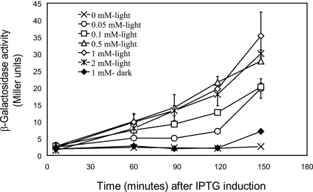
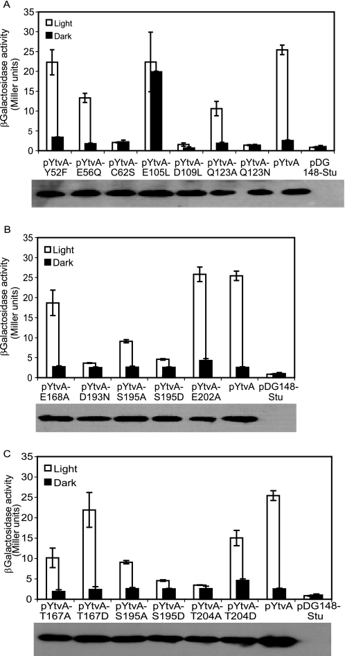
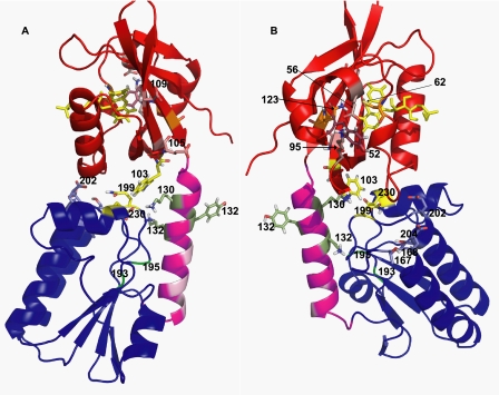
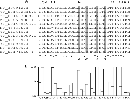
The general stress response of Bacillus subtilis can be activated by stimuli such as the addition of salt or ethanol and with blue light. In the latter response, YtvA activates sigma(B) through a cascade of Rsb proteins, organized in stressosomes. YtvA is composed of an N-terminal LOV (light, oxygen, and voltage) domain and a C-terminal STAS (sulfate transporter and anti-sigma factor) domain and shows light-modulated GTP binding in vitro. Here, we examine the mechanism of YtvA-mediated activation of sigma(B) in vivo with site-directed mutagenesis. Constitutive off and constitutive on mutations have been identified. Disruption of GTP binding in the STAS domain eliminates light activation of sigma(B). In contrast, modification of sites relevant for phosphorylation of STAS domains does not affect the stress response significantly. The data obtained are integrated into a model for the structure of full-length YtvA, which presumably functions as a dimer.


Figures

FIGURE 1.
FIGURE 1.
Optimal conditions for in vivo analysis of YtvA function. Exponentially growing cells of the σB reporter strain that contains Pctc-lacZ, as well as pYtvA, were incubated in the light (open symbols) or dark (closed symbols) with increasing concentrations of IPTG. The x axis shows the time after the addition of 0 (×), 0.05 (○), 0.1 (□), 0.5 (▵), 1 (♢), (♦), and 2 mm (∗) IPTG. The y axis shows the β-galactosidase activity (in Miller units) at the different IPTG concentrations. Error bars indicate S.E. from two duplicate samples.
FIGURE 2.
FIGURE 2.
Effect of mutations in LOV and STAS domains of YtvA on the expression of the σB reporter gene, expressed as Miller units, of the ytvA-overexpressing strains measured 120 min after IPTG addition. In each panel, wild type YtvA and an empty vector (pDG148-Stu) control were also included. Inset of each panel: Western blot analysis of the level of overexpression of each mutant protein. A, mutations in LOV domain as follows: flavin-binding pocket, C62S, Q123A, and Q123N; central β-sheet, E105L and D109L; conserved salt bridge and its surroundings, E56Q and Y52F. B, mutations in STAS domain as follows: GTP binding, D193N, S195A, and S195D; negative charged residues, E168A and E202A. C, mutations in potential phosphorylation sites of STAS domain as follows: T167A, T167D, S195A, S195D, T204A, and T204D.
FIGURE 3.
FIGURE 3.
Ribbon representation of the dark state of the full-length YtvA. Parts A and B represent two views of YtvA, before and after 120° rotation around its longest axis. Numbers indicate the position of the relevant residues. Red, LOV21–125. Magenta, linker region 126–147. Dark blue, STAS148–261. Light red, charged amino acids in the central β-sheet of the LOV domain. Light magenta, hydrophobic residues in the linker region. Bright green, potential GTP-binding site (DSLG). Green, residues Tyr132 and Lys134. Yellow, Trp130 and FMN in the LOV domain, Lys130 in the linker region, and Gln199 and Glu230 in the STAS domain.
FIGURE 4.
FIGURE 4.
A, alignment of the Jα of LOV-STAS containing proteins. Gray boxes, amino acids with hydrophobic and side chains. Letters a and d indicate hydrophobic side chains and correspond to the repeated heptapeptide motif, characteristic for coiled-coil conformation (a, b, c, d, e, f, and g) (46). B, bar graph that represents the average hydropathy of the residues in the sequence alignment. The legend at the x axis indicates a conserved sequence. GenBankTM numbers are as follows: NP_390912.1, B. subtilis; YP_001422316.1, Bacillus amyloliquefaciens FZB42; YP_001487889.1, Bacillus pumilus SAFR-032, ZP_03056406.1, B. pumilus ATCC 7061; YP_001488493.1, B. pumilus SAFR-032; NP_464326.1, Listeria monocytogenes EGD-e; YP_013419.1, L. monocytogenes str. 4b F2365; YP_002350789.1, L. monocytogenes HCC23; NP_470134.1, Listeria innocua Clip11262; YP_848958.1, Listeria welshimeri serovar 6b str. SLCC5334; NP_691509.1, Oceanobacillus iheyensis HTE831; ZP_02171519.1, Bacillus selenitireducens MLS10.

References

    1. Huala E., Oeller P. W., Liscum E., Han I. S., Larsen E., Briggs W. R. (1997) Science 278, 2120–2123 - PubMed
    1. Taylor B. L., Zhulin I. B. (1999) Microbiol. Mol. Biol. Rev. 63, 479–506 - PMC - PubMed
    1. Pellequer J. L., Wager-Smith K. A., Kay S. A., Getzoff E. D. (1998) Proc. Natl. Acad. Sci. U.S.A. 95, 5884–5890 - PMC - PubMed
    1. Christie J. M., Reymond P., Powell G. K., Bernasconi P., Raibekas A. A., Liscum E., Briggs W. R. (1998) Science 282, 1698–1701 - PubMed
    1. Losi A. (2007) Photochem. Photobiol. 83, 1283–1300 - PubMed

LinkOut - more resources