. 2009 Aug 3;206(8):1717-25.
doi: 10.1084/jem.20082492. Epub 2009 Jul 6.

Blockade of CTLA-4 on both effector and regulatory T cell compartments contributes to the antitumor activity of anti-CTLA-4 antibodies

Affiliations

Blockade of CTLA-4 on both effector and regulatory T cell compartments contributes to the antitumor activity of anti-CTLA-4 antibodies

Karl S Peggs et al. J Exp Med. .

Abstract

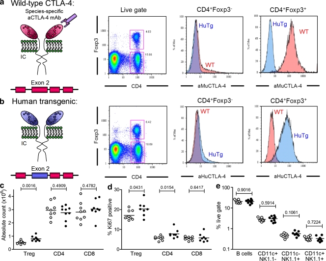
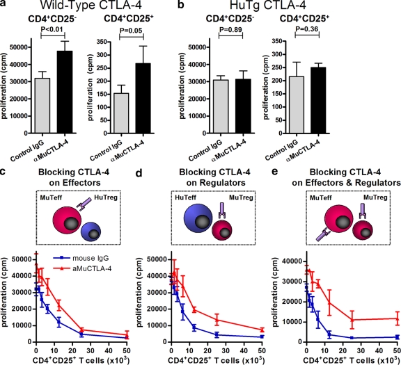
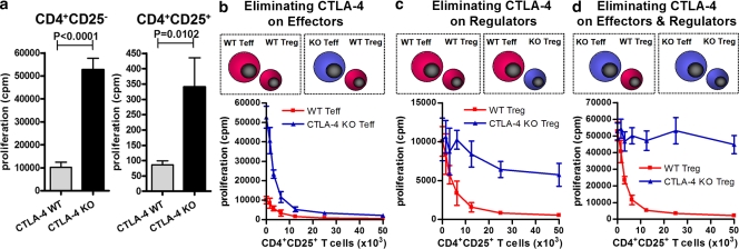
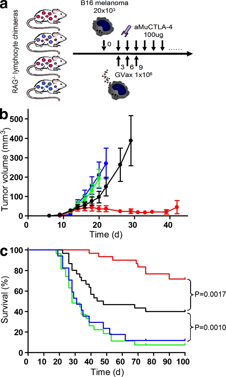
Cytotoxic T lymphocyte-associated antigen 4 (CTLA-4) is a critical negative regulator of immune responses. Uniquely among known inhibitory receptors, its genetic ablation results in a fulminating and fatal lymphoproliferative disorder. This central regulatory role led to the development of antibodies designed to block CTLA-4 activity in vivo, aiming to enhance immune responses against cancer. Despite their preclinical efficacy and promising clinical activity against late stage metastatic melanoma, the critical cellular targets for their activity remains unclear. In particular, debate has focused on whether the effector T cell (T(eff)) or regulatory T cell (T reg cell) compartment is the primary target of antibody-mediated blockade. We developed a mouse expressing human instead of mouse CTLA-4, allowing us to evaluate the independent contributions of CTLA-4 blockade of each T cell compartment during cancer immunotherapy in an in vivo model of mouse melanoma. The data show that although blockade on effector cells significantly improves tumor protection, unicompartmental blockade on regulatory cells completely fails to enhance antitumor responses. However, concomitant blockade of both compartments leads to a synergistic effect and maximal antitumor activity. We conclude that the combination of direct enhancement of T(eff) cell function and concomitant inhibition of T reg cell activity through blockade of CTLA-4 on both cell types is essential for mediating the full therapeutic effects of anti-CTLA-4 antibodies during cancer immunotherapy.


Figures

Figure 1.
Figure 1.
Functional replacement of mouse CTLA-4 with the human CTLA-4 gene in vivo. (a and b) Flow cytometric analysis of intracellular Foxp3 and CTLA-4 in freshly isolated LN CD4+ T cells from WT and HuTg mice, labeled with aMUCTLA-4 (a) and aHuCTLA-4 (b). (c) Absolute CD4+Foxp3+, CD4+Foxp3, and CD8+ T cell counts from age-matched WT (empty circles) and HuTg mice (filled circles; 8–10-mo-old mice; n = 8–9 per group). (d) Frequency of Ki67-expressing CD4+Foxp3+, CD4+Foxp3, and CD8+ T cells in LN of WT (empty circles) and HuTg mice (filled circles). (e) Absolute cell counts for B cell, NK cell, and dendritic cell populations in LN of WT (empty circles) and HuTg mice (filled circles). Data represent three independent experiments. Horizontal bars in c–e represent mean values.
Figure 2.
Figure 2.
Unicompartmental blockade with aMuCTLA-4 during in vitro suppressor assays suggests both cell autonomous and non–cell autonomous activities of CTLA-4. Suppressor assays were performed using mixtures of WT and HuTg cells. (a and b) Proliferation of 50,000 purified WT or HuTg CD4+CD25 and CD4+CD25+ T cells in response to T cell–depleted splenocytes and anti-CD3 and in the presence of control IgG or aMuCTLA-4, confirming the species specificity of the CTLA-4 blockade. (c) Unicompartmental blockade of 50,000 WT CD4+CD25 T cells with aMuCTLA-4, compared with control IgG during in vitro suppression by addition of increasing numbers of HuTg CD4+CD25+ T reg cells. (d) Unicompartmental blockade of WT CD4+CD25+ T reg cells with aMuCTLA-4, compared with control IgG during in vitro suppression of 50,000 HuTg CD4+CD25 T cells. (e) Bicompartmental blockade of both WT CD4+CD25 Teff and CD4+CD25+ T reg cells with aMuCTLA-4, compared with control IgG during in vitro suppressor assays. Data in a–e represent three or more independent experiments, and in each group replicates were performed as quintuplets. Error bars indicate SD.
Figure 3.
Figure 3.
CTLA-4−/− T reg cell display diminished regulatory activity during in vitro suppressor assays. Suppressor assays were performed using mixtures of WT and CTLA-4−/− cells. (a) Proliferation of WT and CTLA-4−/− CD4+CD25 and CD4+CD25+ T cells in response to T cell–depleted splenocytes and anti-CD3, demonstrating cell autonomous inhibitory function of CTLA-4 in the proliferation of Teff and T reg cells. (b) Suppression of 50,000 WT or CTLA-4−/− CD4+CD25 Teff cells by increasing numbers of WT CD4+CD25+ T reg cells. (c) Suppression of 50,000 WT CD4+CD25 Teff cells by increasing numbers of WT or CTLA-4−/− CD4+CD25+ T reg cells. (d) Suppression of 50,000 CTLA-4−/− CD4+CD25 T cells by WT or CTLA-4−/− CD4+CD25+ T reg cells. Data in a–d represent three independent experiments, and in each group replicates were performed as quintuplets. Error bars indicate SD.
Figure 4.
Figure 4.
CTLA-4 blockade of effector and regulatory compartments synergizes to produce maximal antitumor activity. (a) Experimental design. RAG2-deficient mice were reconstituted with four combinations of WT and HuTg T reg and non–T reg (CD4+ and CD8+) cells. 10 wk later, they were challenged with 20 × 103 B16/BL6 melanoma cells intradermally. From day 3, they received combination immunotherapy with Gvax and aMuCTLA-4. (b) Tumor growth curves for mice reconstituted with WT Teff and T reg cells (red), HuTg Teff and WT T reg cells (green), WT Teff and HuTg T reg cells (black), and HuTg Teff and T reg cells (blue; n = 10 per group). Error bars indicate SEM. Data represent three independent experiments. (c) Survival curves with color coding as in b. Mice were euthanized when tumor volume exceeded 350 mm3. Pooled results from three independent experiments are shown.

References

    1. Bachmann M.F., Kohler G., Ecabert B., Mak T.W., Kopf M. 1999. Cutting edge: lymphoproliferative disease in the absence of CTLA-4 is not T cell autonomous.J. Immunol. 163:1128–1131 - PubMed
    1. Bachmann M.F., Gallimore A., Jones E., Ecabert B., Acha-Orbea H., Kopf M. 2001. Normal pathogen-specific immune responses mounted by CTLA-4-deficient T cells: a paradigm reconsidered.Eur. J. Immunol. 31:450–458 - PubMed
    1. Bodor J., Fehervari Z., Diamond B., Sakaguchi S. 2007. ICER/CREM-mediated transcriptional attenuation of IL-2 and its role in suppression by regulatory T cells.Eur. J. Immunol. 37:884–895 - PubMed
    1. Butte M.J., Keir M.E., Phamduy T.B., Sharpe A.H., Freeman G.J. 2007. Programmed death-1 ligand 1 interacts specifically with the B7-1 costimulatory molecule to inhibit T cell responses.Immunity. 27:111–122 - PMC - PubMed
    1. Carreno B.M., Bennett F., Chau T.A., Ling V., Luxenberg D., Jussif J., Baroja M.L., Madrenas J. 2000. CTLA-4 (CD152) can inhibit T cell activation by two different mechanisms depending on its level of cell surface expression.J. Immunol. 165:1352–1356 - PubMed

Publication types

MeSH terms