. 2009 Jul 21;106(29):12162-7.
doi: 10.1073/pnas.0901915106. Epub 2009 Jul 6.

Optogenetic control of epileptiform activity

Affiliations

Optogenetic control of epileptiform activity

Jan Tønnesen et al. Proc Natl Acad Sci U S A. .

Abstract

The optogenetic approach to gain control over neuronal excitability both in vitro and in vivo has emerged as a fascinating scientific tool to explore neuronal networks, but it also opens possibilities for developing novel treatment strategies for neurologic conditions. We have explored whether such an optogenetic approach using the light-driven halorhodopsin chloride pump from Natronomonas pharaonis (NpHR), modified for mammalian CNS expression to hyperpolarize central neurons, may inhibit excessive hyperexcitability and epileptiform activity. We show that a lentiviral vector containing the NpHR gene under the calcium/calmodulin-dependent protein kinase IIalpha promoter transduces principal cells of the hippocampus and cortex and hyperpolarizes these cells, preventing generation of action potentials and epileptiform activity during optical stimulation. This study proves a principle, that selective hyperpolarization of principal cortical neurons by NpHR is sufficient to curtail paroxysmal activity in transduced neurons and can inhibit stimulation train-induced bursting in hippocampal organotypic slice cultures, which represents a model tissue of pharmacoresistant epilepsy. This study demonstrates that the optogenetic approach may prove useful for controlling epileptiform activity and opens a future perspective to develop it into a strategy to treat epilepsy.


Conflict of interest statement

The authors declare no conflict of interest.

Figures

Fig. 1.
Fig. 1.
NpHR expression in organotypic hippocampal slice cultures. (A) Epifluorescence microscopy image showing EYFP immunoreactivity in several hippocampal regions after LV-NpHR-EYFP-pCaMKIIα vector–mediated transduction. (Scale bar, 200 μm.) Boxed areas are shown magnified on the right. EYFP-labeled granule cells (B) in the DG, and EYFP-labeled pyramidal neurons in the CA1 (C) and CA3 (D) region of the hippocampus. (E) Retrospective identification of biocytin-labeled pyramidal cell in CA3 after whole-cell patch-clamp recording. Yellow indicates merged double-labeling of EYFP (FITC; green) and biocytin (Cy3; red). (Scale bar in B, 100 μm for B–E.)
Fig. 2.
Fig. 2.
I-V relationship in NpHR-transduced CA1 and CA3 pyramidal neurons. (A and C) Voltage responses in NpHR-transduced CA1 (A) and CA3 (C) pyramidal neurons during current step injections of 500-msec duration. (B) Averaged I-V relationship of NpHR-transduced (n = 12 cells) and nontransduced (n = 11 cells) CA1 pyramidal neurons during all steps of current injection. Note that no light is applied during these experiments. (D) Averaged I-V relationship curves in NpHR-transduced (n = 9 cells) as compared with nontransduced (n = 10 cells) CA3 pyramidal neurons during current step injections. Note significant differences at hyperpolarizing steps. *P < 0.05.
Fig. 3.
Fig. 3.
Orange-light illumination hyperpolarizes NpHR-transduced pyramidal cells and suppresses the generation of action potentials. (A and C) Initial and steady voltage hyperpolarizations by NpHR activation in CA1 (A) and CA3 pyramidal (C) neurons. (B and D) Inhibition of current injection-induced action potentials by orange-light illumination in CA1 (B) and CA3 (D) pyramidal neurons. Bars illustrate time of light illumination.
Fig. 4.
Fig. 4.
The reversal potential of GABAA receptor–mediated IPSCs is unchanged by sustained orange-light activation of transgene NpHR. (A) Orange-light illumination hyperpolarizes NpHR-transduced EYFP-expressing granule cells recorded using perforated whole-cell patch-clamp configuration. (B) Voltage-clamp of the same cell as in A during application of 10-mV voltage steps (from −50 to −90 mV) combined with stimulation of afferent fibers generating monosynaptic IPSCs (indicated in dashed box and magnified on the right). The stimulation protocol was repeated during orange-light illumination of the organotypic slice culture. (C) After perforated whole-cell patch-clamp recording, the cell membrane was ruptured by negative pressure, allowing biocytin to diffuse into the cell from the attached pipette, and the recorded cell in A was retrospectively stained for EYFP and biocytin double-labeling (superimposed as yellow). (Scale bar, = 50 μm; applies to all images.) (D) Average amplitudes of evoked IPSCs plotted against the corresponding voltage step during no light and orange-light illumination (n = 5 cells).
Fig. 5.
Fig. 5.
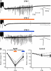
STIB in CA3 is strongly attenuated by orange-light activation of transgene NpHR in organotypic hippocampal cultures. (A) Recordings of 3 consecutive STIB stimulations, with orange-light illumination on second stimulation, in NpHR-transduced slices. Insets: Magnification of traces showing epileptiform bursts after STIB stimulation. Scale bars apply for all traces. (B) Relative changes in STIB duration of individual recordings (dotted lines and open circles) and their average values (mean ± SEM; solid line and squares) for NpHR and nontransduced control slices, respectively, during 3 consecutive STIB. **P < 0.01; ***P < 0.001.
Fig. 6.
Fig. 6.
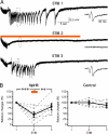
STIB in CA1 is strongly attenuated by orange-light activation of transgene NpHR in organotypic hippocampal cultures. (A) Recordings of 3 consecutive STIB stimulations, with orange-light illumination on second stimulation, in NpHR-transduced slices. Insets: Magnification of traces showing spikes during STIB. Scale bars apply for all traces. (B) Relative changes in STIB duration of individual recordings (dotted lines and open circles) and their average values (mean ± SEM; solid line and filled squares) for NpHR and nontransduced control slices, respectively, during 3 consecutive STIB. At STIB2, orange light is applied to NpHR-transduced slices as indicated by orange bar. At STIB3, blue light is applied to NpHR-transduced slices as indicated by blue bar. ***P < 0.001.
Fig. 7.
Fig. 7.
PDS occurrence during STIB in NpHR-transduced CA3 pyramidal neurons during STIB is strongly inhibited by orange-light stimulation. (A) PDS in CA3 pyramidal cell recorded by whole-cell patch-clamp during 3 consecutive STIB. (B) Light activation of NpHR (shown by orange bar) at STIB2 reduces the duration of PDS occurrence without changing number of action potentials per PDS. (C) Relative changes in duration of PDS occurrence recorded in individual neurons (indicated by dotted lines and open circles) and average values, indicated by solid line (mean ± SEM), for NpHR-transduced and nontransduced CA3 pyramidal neurons. (D) Repeated orange-light illumination (intermediated by control stimulations without light exposure) repeatedly inhibits STIB-generated PDS occurrence by activation of NpHR. **P < 0.01; ***P < 0.001.

References

    1. Falb M, et al. Living with two extremes: Conclusions from the genome sequence of Natronomonas pharaonis. Genome Res. 2005;15:1336–1343. - PMC - PubMed
    1. Kolbe M, Besir H, Essen LO, Oesterhelt D. Structure of the light-driven chloride pump halorhodopsin at 1.8 A resolution. Science. 2000;288:1390–1396. - PubMed
    1. Zhang F, Aravanis AM, Adamantidis A, de Lecea L, Deisseroth K. Circuit-breakers: Optical technologies for probing neural signals and systems. Nat Rev Neurosci. 2007;8:577–581. - PubMed
    1. Zhang F, et al. Multimodal fast optical interrogation of neural circuitry. Nature. 2007;446:633–639. - PubMed
    1. Banerjee PN, Hauser WA. Incidence and prevalence. In: Engel JJ, Pedley T, editors. Epilepsy: a comprehensive textbook. 2nd Ed. Baltimore: Wolters Kluwer/Lippincott Williams & Wilkins; 2008. pp. 45–56.

Publication types

MeSH terms