. 2009 Jul 14;106(28):11691-6.
doi: 10.1073/pnas.0902426106. Epub 2009 Jul 6.

NPAS3 is a trachealess homolog critical for lung development and homeostasis

Affiliations

NPAS3 is a trachealess homolog critical for lung development and homeostasis

Shutang Zhou et al. Proc Natl Acad Sci U S A. .

Erratum in

Abstract

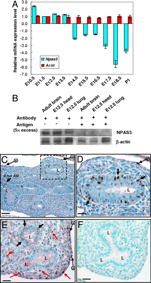
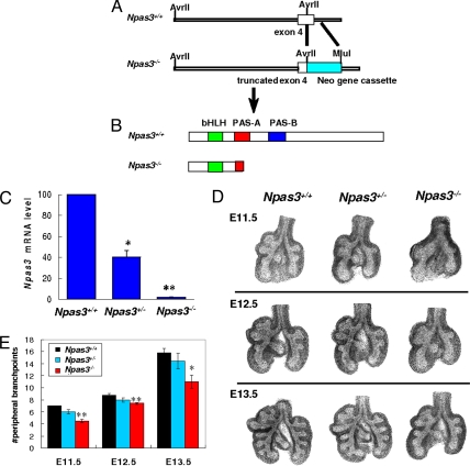
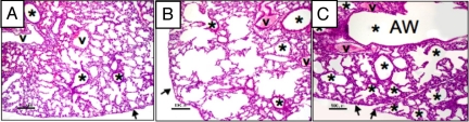
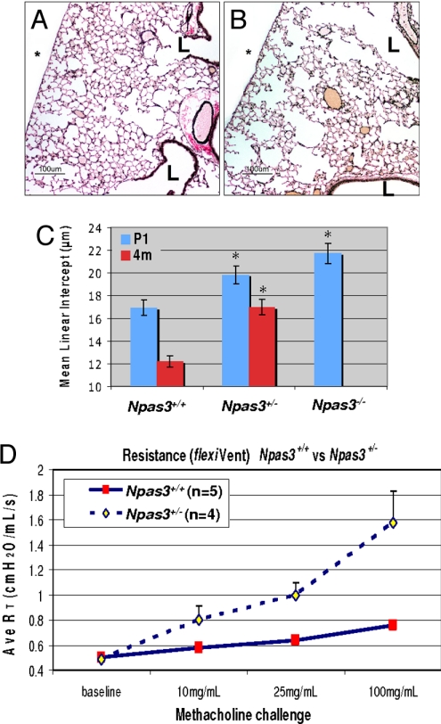
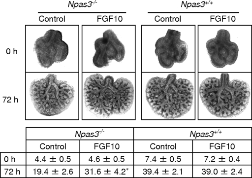
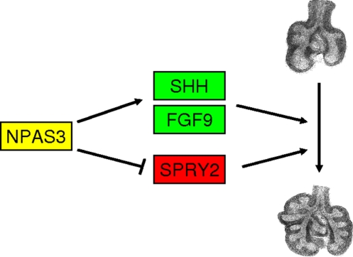
Trachealess (Trh) is a PAS domain transcription factor regulating Drosophila tracheogenesis. No other Trh homolog has been associated with a respiratory phenotype. Seeking homolog(s) regulating lung development, we screened murine genomic DNA using trh oligonucleotides, identifying only Npas3. Npas3 mRNA peaks in lung from E10.5 to E13.5, verified by sequencing, with immunostaining in airway epithelial cells. Npas3-null mice have reduced lung branching morphogenesis but are viable prenatally. Npas3-null newborns die in respiratory distress, with diminished alveolarization, decreased Shh, Fgf9, Fgf10, and Bmp4 mRNAs, and increased Spry2, consistent with reduced FGF signaling. Exogenous FGF10 rescues branching morphogenesis in Npas3-null lungs. In promoter reporter assays, NPAS3 directly upregulates Shh and represses Spry2. Npas3(+/-) mice have a milder lung phenotype, surviving postnatally, but develop emphysema and airways hyperreactivity. Therefore, absence of a developmentally expressed transcription factor can alter downstream gene expression and multiple signaling pathways in organogenesis. NPAS3 haploinsufficiency may also lead to emphysema and asthma.


Conflict of interest statement

The authors declare no conflict of interest.

Figures

Fig. 1.
Fig. 1.
Developmental expression of Npas3 mRNA and protein in murine lungs. (A) QRT-PCR analysis of Npas3 and Arnt mRNA in lungs from E10.5 to P1. (B) Western blot analysis of NPAS3 in E12.5 lungs, using adult brains and E12.5 heads as positive controls, and preabsorbing NPAS3 antibody with excess antigen for specificity. (C–F) NPAS3 immunohistochemistry in E13.5 (C and D) and E15.5 (E and F) lungs. Some NPAS3-positive epithelial nuclei (black arrows), mesothelial cells (long black arrows with asterisks), and mesenchymal cells (red arrows) are indicated. D is the higher magnification of dashed line box in C; F is a section immediately serial to E, running in parallel using antigen-preabsorbed NPAS3 antibody. L, airway lumen; Prox. AW, proximal airway. (Scale bars: C, 50 μm; D–F, 25 μm.)
Fig. 2.
Fig. 2.
Npas3 targeting and branching morphogenesis in Npas3-deficient lungs. (A and B) To generate Npas3-null mice, a 1.4-kb DNA fragment in exon 4 and intron 4 of murine Npas3 gene was replaced by Neo gene cassette, leading to a frameshift mutation and premature termination. (C) QRT-PCR analysis of Npas3 mRNA from Npas3-deficient vs. WT littermate lungs at E12.5. *P < 0.01, ** P < 0.001, n = 4. (D) Representative WT vs. Npas3-deficient lung buds at E11.5, E12.5, and E13.5. (E) Pooled results of numbers of peripheral branch points in lung buds, with 4–5 litters per timepoint. Branching of Npas3−/− lung buds significantly reduced compared with Npas3+/+ littermates. *P < 0.05, **P < 0.01.
Fig. 3.
Fig. 3.
Histopathological features of Npas3-deficient lungs at P1. Lung histopathology at P1 for (A) WT, (B) Npas3-Het, and (C) Npas3-null littermates. Compared with WT, Npas3-deficient lungs have dilated airways with few alveoli. Npas3-Hets have defects in the primitive alveolar parenchyma alternated with segments of more normal alveoli (saccular stage) and many airways extend near the pleura (arrows). Npas3-null pups have reduced alveolar parenchyma. The conducting airways are massively dilated, similar to bronchiectasis. v, blood vessel; *, airway (AW). (Scale bar: 100 μm.)
Fig. 4.
Fig. 4.
Lung histopathology of adult Npas3-deficient mice. (A and B) Lung histopathology from WT (A) and Npas3-Het littermates (B) at 4 months (4 mo) of age (Voerhoff's elastic tissue stain). L, airway lumen; *, pleural surface; bar, 100 μm. (C) Mean linear intercepts of lungs from P1 and 4-mo mice (3 litters each): *P < 0.0001 compared with WT. Note that there were no 4-mo-old Npas3-null mice because pups rarely survived beyond 48 h.
Fig. 5.
Fig. 5.
FGF10 restores branching of Npas3-null embryonic lungs in vitro. Littermate Npas3+/+ and Npas3−/− E11.5 lungs were cultured in growth medium alone (control) or plus 500 ng/mL recombinant human FGF10 for 72 h. (Upper) Representative lung buds cultured in medium or medium plus recombinant FGF10 at 0 h and 72 h. (Lower) Pooled data of lung peripheral branch-point number (mean ± SD) at 0 h and 72 h. Asterisk (*) indicates branching of Npas3−/− lung buds was significantly increased with FGF10 at 72 h (P < 0.01, n = 5), but was less than littermate WT control (P < 0.05, n = 5).
Fig. 6.
Fig. 6.
Working hypothesis. Schematic drawing of how NPAS3 might function in epithelial and mesenchymal cells in developing lungs. NPAS3 regulates transcription of Shh and Fgf9, which would lead to increased SHH signaling via PATCHED with activation of Gli2 and the cascade of genes activated by SHH signaling. NPAS3 activation of FGF signaling includes FGF9 and FGF10, which reciprocally activate both epithelial and mesenchymal cells. FGFR2 signaling is inhibited by SPRY2, but NPAS3 represses Spry2 expression, and thus facilitates FGF signaling during lung development.

Comment in

  • Findings of Research Misconduct.
    [No authors listed] [No authors listed] Fed Regist. 2019 Nov 7;84(216):60097-60098. Fed Regist. 2019. PMID: 37547121 Free PMC article. No abstract available.

References

    1. Manning G, Krasnow MA. Development of Drosophila Melanogaster. Cold Spring Harbor, NY: Cold Spring Harbor Lab Press; 1993. Development of the Drosophila tracheal system; pp. 609–685.
    1. Affolter M, et al. Tube or not tube: Remodeling epithelial tissues by branching morphogenesis. Dev Cell. 2003;4:11–18. - PubMed
    1. Wilk R, Weizman I, Shilo B-Z. Trachealess encodes a bHLH-PAS protein that is an inducer of tracheal cell fates in Drosophila. Genes Dev. 1996;10:93–102. - PubMed
    1. Metzger RJ, Krasnow MA. Genetic control of branching morphogenesis. Science. 1999;284:1635–1639. - PubMed
    1. Kewley RJ, Whitelaw ML, Chapman-Smith A. The mammalian basic helix-loop-helix/PAS family of transcriptional regulators. Int J Biochem Cell Biol. 2004;36:189–204. - PubMed

Publication types

MeSH terms