. 2009 Sep 4;284(36):23972-9.
doi: 10.1074/jbc.M109.010199. Epub 2009 Jul 6.

Role of dimerization in the catalytic properties of the Escherichia coli disulfide isomerase DsbC

Affiliations

Role of dimerization in the catalytic properties of the Escherichia coli disulfide isomerase DsbC

Silvia A Arredondo et al. J Biol Chem. .

Abstract

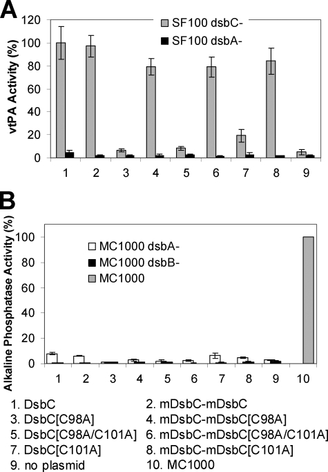
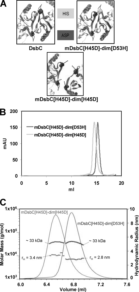
The bacterial protein-disulfide isomerase DsbC is a homodimeric V-shaped enzyme that consists of a dimerization domain, two alpha-helical linkers, and two opposing thioredoxin fold catalytic domains. The functional significance of the two catalytic domains of DsbC is not well understood yet. We have engineered heterodimer-like DsbC derivatives covalently linked via (Gly(3)-Ser) flexible linkers. We either inactivated one of the catalytic sites (CGYC), or entirely removed one of the catalytic domains while maintaining the putative binding area intact. Variants having a single active catalytic site display significant levels of isomerase activity. Furthermore, mDsbC[H45D]-dim[D53H], a DsbC variant lacking an entire catalytic domain but with an intact dimerization domain, also showed isomerase activity, albeit at lower levels. In addition, the absence of the catalytic domain allowed this protein to catalyze in vivo oxidation. Our results reveal that two catalytic domains in DsbC are not essential for disulfide bond isomerization and that a determining feature in isomerization is the availability of a substrate binding domain.


Figures

FIGURE 1.
FIGURE 1.
A, protein structure of DsbC, and molecular models of mDsbC-mDsbC and the single active site covalently linked mutants. Dimerization domains are shown in gray, thioredoxin domains in black, and the active sites in white. B, gel filtration FPLC of DsbC and linked variants. Purified proteins were run on a SuperdexTM 200 column in PBS, 10% glycerol buffer.
FIGURE 2.
FIGURE 2.
In vivo activity of single active site mutants. A, yield of active vtPA in dsbC (gray bars) or dsbA (black bars) cells. E. coli SF100 ΔdegP ΔdsbC and SF100 ΔdegP dsbA::kan (respectively) were transformed with pTrcStIIvtPA and pBAD33 encoding the respective DsbC derivatives. Protein synthesis was induced as described under “Experimental Procedures,” and the yield of active vtPA at 3 h after induction was determined. Relative activities were obtained by dividing the ΔA405 of each strain by the ΔA405 of the strain expressing wild type DsbC. B, PhoA activity in E. coli MC1000 dsbA::kan (white bars) and MC1000 dsbB::kan (black bars). The alkaline phosphatase activity in the parental isogenic strain MC1000 is shown by the gray bar.
FIGURE 3.
FIGURE 3.
A, molecular model of mDsbC-dim. Dimerization domains are shown in gray, thioredoxin domain in black, and catalytic site in white. B, gel filtration FPLC of mDsbC-dim as compared with DsbC. Purified proteins were run on a SuperdexTM 200 column in PBS, 10% glycerol buffer. C, MALS measurement of the molar masses of the components of mDsbC-dim together with their hydrodynamic radii. The data show monomeric, dimeric, and tetrameric states. The relative concentrations were determined by the refractive index differences.
FIGURE 4.
FIGURE 4.
A, crystal structure of DsbC at the dimerization interface is shown. Residues His45 and Asp53 interact to establish salt bridges. In mDsbC[H45D]-dim[D53H], His45 is replaced by Asp in the first dimerization domain and Asp53 is replaced by His in the second dimerization domain to disrupt interdimerization but promote intradimerization. In mDsbC[H45D]-dim[H45D], residue His45 in each dimerization domain is replaced by Asp to fully disrupt dimerization. B, gel filtration FPLC of mDsbC[H45D]-dim[D53H] and mDsbC[H45D]-dim[H45D]. Purified proteins were run on a SuperdexTM 200 column in PBS-10% glycerol. C, MALS measurement of the molar masses of mDsbC[H45D]-dim[D53H] and mDsbC[H45D]-dim[H45D]. The relative concentrations are shown as determined by the refractive index differences. Both mutants have similar molar masses but elute at different volumes in the order predicted by their hydrodynamic radii.
FIGURE 5.
FIGURE 5.
In vivo activity of single catalytic-domain mutants. A, yield of active vtPA in SF100 ΔdegP ΔdsbC (gray bars) or SF100 ΔdegP dsbA::kan (black bars) cells was determined as described under “Experimental Procedures.” B, PhoA activity in E. coli MC1000 dsbA::kan (white bars) and MC1000 dsbB::kan (black bars). The alkaline phosphatase activity in the parental isogenic strain MC1000 is shown by the gray bar.

Similar articles

Cited by

References

    1. Gleiter S., Bardwell J. C. (2008) Biochim. Biophys. Acta 1783, 530–534 - PMC - PubMed
    1. Ito K., Inaba K. (2008) Curr. Opin. Struct. Biol. 18, 450–458 - PubMed
    1. Kadokura H., Katzen F., Beckwith J. (2003) Annu. Rev. Biochem. 72, 111–135 - PubMed
    1. Zapun A., Bardwell J. C., Creighton T. E. (1993) Biochemistry 32, 5083–5092 - PubMed
    1. Grauschopf U., Fritz A., Glockshuber R. (2003) EMBO J. 22, 3503–3513 - PMC - PubMed

Publication types

MeSH terms

Substances

LinkOut - more resources