. 2009 Oct;66(4):448-54.
doi: 10.1203/PDR.0b013e3181b45565.

Characterization and culture of fetal rhesus monkey renal cortical cells

Affiliations

Characterization and culture of fetal rhesus monkey renal cortical cells

Alyssa C Leapley et al. Pediatr Res. 2009 Oct.

Abstract

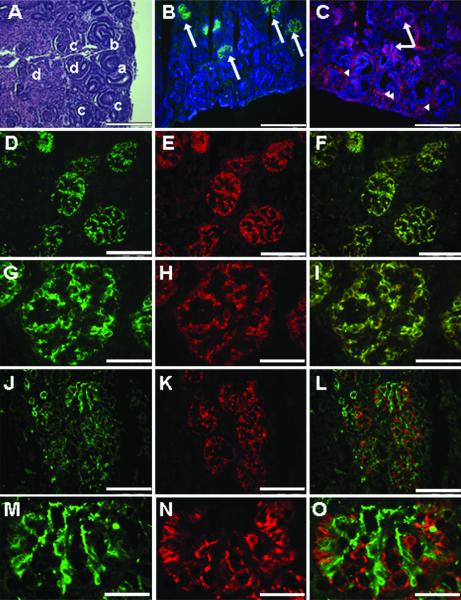
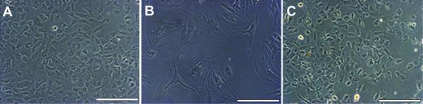
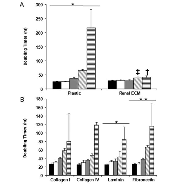
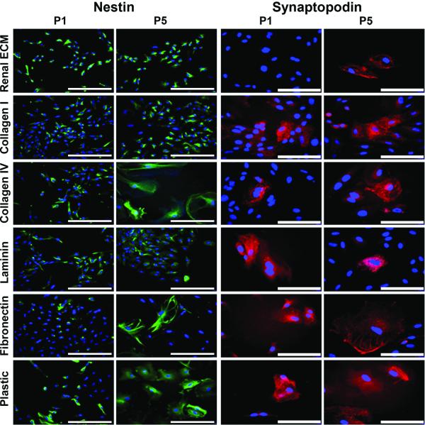
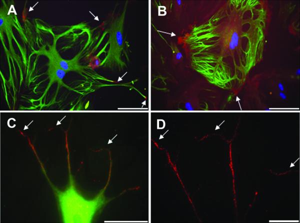
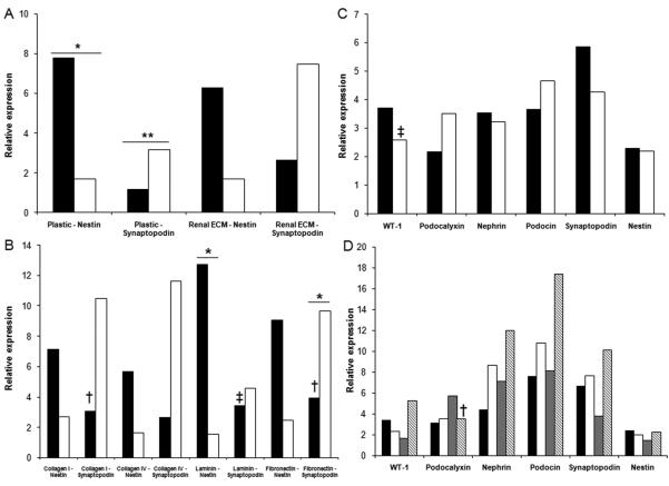
The renal glomerulus is composed of endothelial and mesangial cells with podocytes contributing to glomerular filtration. Podocyte damage is associated with renal disorders, thus there is interest in these cells for regenerative medicine. These studies investigated the use of extracellular matrix (ECM) to grow third trimester fetal monkey renal cortical cells and to assess mature podocytes in culture. Immunohistochemistry provided a profile of podocyte differentiation with metanephric mesenchyme and developing podocytes nestin positive and synaptopodin negative, whereas mature podocytes were positive for both markers. Primary cell cultures devoid of mature podocytes were established on plastic and renal ECM. A cell population (nestin+/synatopodin-) cultured on renal ECM showed greater proliferative potential compared with plastic with limited podocytes developing in culture over time. Further investigation of individual components of ECM (laminin, fibronectin, collagen I, or collagen IV) indicated that collagen I supported the greatest proliferation similar to renal ECM, whereas a greater number of mature podocytes (nestin+/synaptopodin+) were observed on fibronectin. These results suggest that (1) culture of fetal monkey podocytes can be accomplished, (2) renal ECM and collagen I can support renal cortical cells in vitro, which may recapitulate the developing kidney in vivo, and (3) fibronectin can support podocyte differentiation in vitro.


Figures

Figure 1
Figure 1. Renal morphology and immunohistochemistry - Early third trimester fetal monkey kidney and nephrogenic zone
(A) The fetal monkey kidney in the early third trimester shows a clear nephrogenic zone in the cortical region with the ureteric bud (arrowhead) and the metenephric mesenchyme (arrow) evident. The four stages of developing glomeruli are also present and represent the vesicle (a), S-shape (b), capillary (c), and mature (d) glomerulus. Hematoxylin and eosin (H&E). (B-C) The nephrogenic zone is shown with the nuclei stained with DAPI (blue), metanephric mesenchyme (arrowheads), developing podocytes, and mature podocytes (arrows) positive for synaptopodin (green) and nestin (red). (D-I) Mature podocytes in terminally differentiated glomeruli are positive for nestin (red) and synaptopodin (green). (J-O) The endothelial marker CD31 (green) is not evident with the nestin positive cells (red) in these specimens (A-C, 20x, bar = 100 μm; D-F, 40x, bar = 50 μm; G-I, 100x, bar = 20 μm; J-L, 40x, bar = 50 μm; M-O, 100x, bar = 20 μm).
Figure 2
Figure 2. Morphology of cultured renal cortical cells
(A) Through passage 4, all substrates studied showed cells with the “cobblestone” appearance suggestive of undifferentiated podocytes. (B) At passage 5, cultures contained cells that had a differentiated podocyte cell morphology with arborization, processes extended from cell bodies, and a large cytoplasmic to nuclear volume ratio. (C) Renal ECM contained cells with an undifferentiated podocyte morphology through all passages (1 through 5) (20x, bar = 100 μm).
Figure 3
Figure 3. Population doubling times - Renal cortical cells on renal ECM
(A-B) (black - P1, white - P2, gray - P3, diagonal hatched - P4, vertical hatched - P5) The Y-axis represents population doubling times (h). The population doubling times suggest that in the early passages, the substrate did not alter the growth capabilities of the cells. For passages 4 and 5, renal ECM was shown to support the optimal population doubling times. Comparisons were made of passage 1 (P1) versus passage 5 (P5) for each substrate; *p<0.05, **p<0.01. Comparisons of renal ECM with plastic are shown at passage 4 (p<0.01), and renal ECM with plastic, fibronectin, and laminin at passage 5 (p<0.05).
Figure 4
Figure 4. Immunocytochemistry - Cultured cortical cells
Cells from passage 1 (P1) grown on each of the substrates included nestin positive cells (green) (20x, bar = 100 μm). Nuclei stained with DAPI (blue). At passage 5 (P5), nestin positive cells decreased on all substrates except renal ECM and collagen I. Cells from P1 cultured on five of six substrates contained very few cells that were synaptopodin positive (red, 40x, bar = 50 μm). None of the cells grown on renal ECM showed evidence of synaptopodin expression. At P5, there was an increase in synaptopodin positive cells suggesting a more mature podocyte phenotype.
Figure 5
Figure 5. Immunocytochemistry - Cultured cortical cells on fibronectin and renal ECM
Cells from passage 5 cultured on fibronectin contained more synaptopodin positive cells (red) (arrow) than those cultured on other substrates. Nuclei stained with DAPI (blue) (A-D). Synaptopodin positive cells were positive for nestin (green) and showed morphology consistent with mature podocytes (A-C). (A-B, 20x, bar = 100 μm; C, 60x, bar = 50 μm; D, 100x, bar = 20 μm).
Figure 6
Figure 6. Quantitative PCR - Synaptopodin and nestin
While synaptopodin expression increased (A), nestin expression decreased (B) in cells grown on all substrates studied (black column - P1, white column - P5). Furthermore, podocyte specific expression was present in cells at P5 (C) (black - plastic, white - renal ECM) (D) (black - collagen I, white - collagen IV, gray - laminin, hatched - fibronectin). Passage 1 (P1) compared to passage 5 (P5) (*p<0.05, **p<0.01). Comparisons of plastic to other substrates (p<0.05, p<0.01).

References

    1. Quaggin SE, Kreidberg JA. Development of the renal glomerulus: good neighbors and good fences. Development. 2008;135:609–620. - PubMed
    1. Saxen L. Organogenesis of the kidney. Cambridge University Press; Cambridge: 1987. pp. 135–142.
    1. Miner JH. Renal basement membrane components. Kidney Int. 1999;56:2016–2024. - PubMed
    1. Haller H, de Groot K, Bahlmann F, Elger M, Fliser D. Stem cells and progenitor cells in renal disease. Kidney Int. 2005;68:1932–1936. - PubMed
    1. Bertelli E, Regoli M, Fonzi L, Occhini R, Mannucci S, Ermini L, Toti P. Nestin expression in adult and developing human kidney. J Histochem Cytochem. 2007;55:411–421. - PubMed

Publication types

MeSH terms