. 2009 Sep 24;28(38):3380-9.
doi: 10.1038/onc.2009.193. Epub 2009 Jul 6.

p21(WAF1) gene promoter is epigenetically silenced by CTIP2 and SUV39H1

Affiliations

p21(WAF1) gene promoter is epigenetically silenced by CTIP2 and SUV39H1

T Cherrier et al. Oncogene. .

Abstract

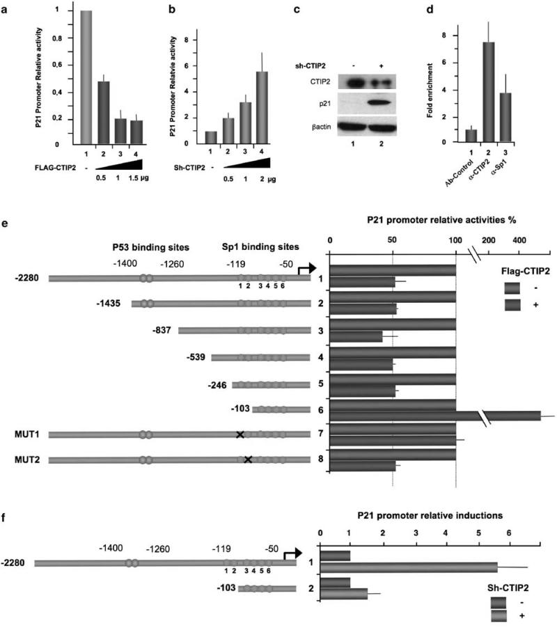
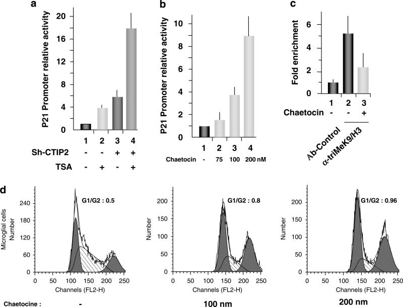
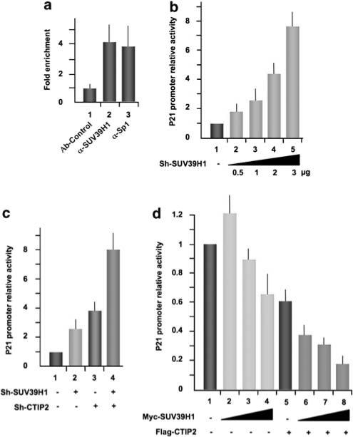
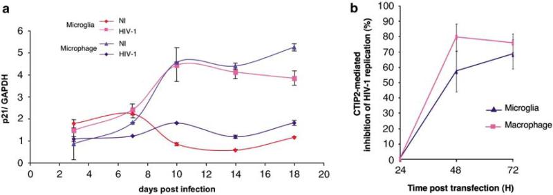
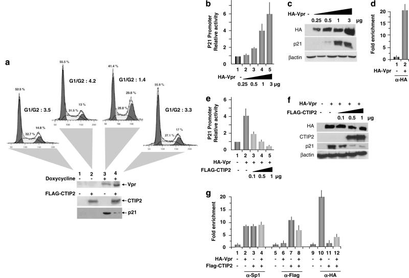
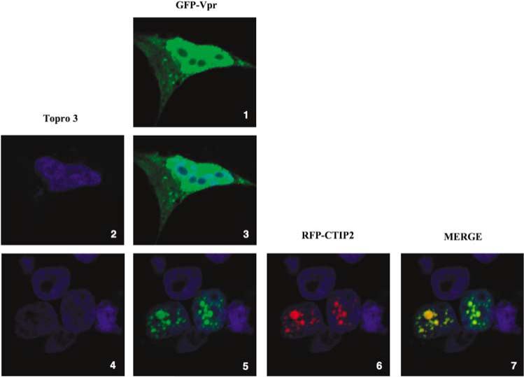
Mainly regulated at the transcriptional level, the cellular cyclin-dependent kinase inhibitor, CDKN1A/p21(WAF1) (p21), is a major cell cycle regulator of the response to DNA damage, senescence and tumor suppression. Here, we report that COUP-TF-interacting protein 2 (CTIP2), recruited to the p21 gene promoter, silenced p21 gene transcription through interactions with histone deacetylases and methyltransferases. Importantly, treatment with the specific SUV39H1 inhibitor, chaetocin, repressed histone H3 lysine 9 trimethylation at the p21 gene promoter, stimulated p21 gene expression and induced cell cycle arrest. In addition, CTIP2 and SUV39H1 were recruited to the silenced p21 gene promoter to cooperatively inhibit p21 gene transcription. Induction of p21(WAF1) gene upon human immunodeficiency virus 1 (HIV-1) infection benefits viral expression in macrophages. Here, we report that CTIP2 further abolishes Vpr-mediated stimulation of p21, thereby indirectly contributing to HIV-1 latency. Altogether, our results suggest that CTIP2 is a constitutive p21 gene suppressor that cooperates with SUV39H1 and histone methylation to silence the p21 gene transcription.


Figures

Figure 1
Figure 1
Associated with the p21 proximal promoter region in vivo, COUP-TF-interacting protein 2 (CTIP2) impairs constitutive p21 gene transcription through the proximal –246/–103 Sp1-binding sites. Microglial cells (a and b) and 293T cells (c) were transfected with the indicated amounts of vectors in the presence of the p21-Luc plasmid. Luciferase activities (a and b) are presented relative to the activity of the p21-LUC cotransfected with the control empty vector. The protein levels (c) were accessed by western blot with the indicated antibodies 48-h post-transfection. (d) To detect association of CTIP2 and Sp1 with the p21 promoter, ChIP (chromatin immunoprecipitation) experiments were performed in microglial cells with the indicated antibodies. Immunoprecipitated DNA was subjected to real-time PCR quantification with primers targeting the endogenous p21 promoter proximal region. Specific enrichments of the p21 promoter in the immunoprecipitated material were normalized to enrichments in non-specific glyceraldehyde 3-phosphate dehydrogenase DNA and are presented relative to the control. (e and f) Microglial cells were transfected with the p21-LUC constructs in the presence or absence of the pFlag-CTIP2 or pShRNA-CTIP2 plasmids, as indicated. Luciferase assays were performed 48-h post-transfection. Results are presented relative to the basal level of each p21-LUC constructs taken as one.
Figure 2
Figure 2
Trichostatin A (TSA) and chaetocin stimulate p21 gene transcription. (a, b) Microglial cells were transfected with the p21-LUC reporter plasmid in the presence or absence of pSuper-shRNA-CTIP2, as indicated. At 24-h post-transfection, cells were treated with 450 μm TSA or the indicated amount of chaetocin for 24 h and assessed for LUC activity. (c) Microglial cells were treated or not with 200 nm of chaetocin and subjected to ChIP (chromatin immunoprecipitation) experiments with anti-H3 trimethylated lysine 9. Immunoprecipitated DNA was subjected to real-time PCR quantification with primers targeting the endogenous p21 promoter proximal region. Specific enrichment of the p21 promoter in the immunoprecipitated material were normalized to enrichments in nonspecific glyceraldehyde 3-phosphate dehydrogenase DNA and presented relative to the control. (d) Microglial cells were treated with the indicated amount of chaetocin for 24 h before being assessed for cell cycle status by flow cytometry.
Figure 3
Figure 3
COUP-TF-interacting protein 2 (CTIP2) cooperates with SUV39H1 to repress p21 gene transcription. (a) ChIP (chromatin immunoprecipitation) experiments were performed in microglial cells with the indicated antibodies. Immunoprecipitated DNA was subjected to real-time PCR quantification with primers targeting the endogenous p21 promoter proximal region. Specific enrichment of the p21 promoter in the immunoprecipitated material were normalized to enrichments in non-specific glyceraldehyde 3-phosphate dehydrogenase DNA and presented relative to the control. (bd) Microglial cells were transfected with the p21-LUC reporter plasmid in the presence or absence of the indicated vectors. LUC assays were performed 48-h post-transfection and results presented relative to the basal level.
Figure 4
Figure 4
As P21 gene expression is upregulated by human immunodeficiency virus 1 (HIV-1) infection, COUP-TF-interacting protein 2 (CTIP2) inhibits HIV-1 replication in human primary macrophages and microglial-like cells. (a) RNAs from HIV-1/Bal-infected cells were extracted and assessed for p21 mRNA expression. Inductions at each time points are presented relative to the control glyceraldehyde 3-phosphate dehydrogenase level. (b) Cells were transfected with HIV-1 pNL4-3 in the presence or absence (control cells) of the Flag-CTIP2 expression vector. HIV-1 reverse transcriptase activity was assessed 24-, 48- and 72-h post-transfection. Inhibition of HIV-1 replication is presented relative to the reverse transcriptase (RT) activity detected with the HIV-1-infected control cells. RT activity in control cells (without Flag-CTIP2) varied between 0 and 197 pg/ml.
Figure 5
Figure 5
COUP-TF-interacting protein 2 (CTIP2) impairs Vpr-mediated stimulation of p21 gene transcription and the resulting cell cycle arrest. (a) 293T cells inducible for HIV-1 Vpr expression, were transfected with the Flag-CTIP2 expression vector or the control vector 24 h before being treated with doxycyclin. Cell cycle status was assessed by flow cytometry. Controls of Vpr, CTIP2 and p21 expressions are presented. Microglial cells (b and e) and 293 T cells (c and f) were transfected with the indicated amounts of expression vectors in the presence of the p21-Luc plasmid. Luciferase activities (b and e) are presented relative to the activity of the p21-LUC complemented by the control empty vector. Protein levels (c and f) were assessed by western blot using the indicated antibodies 48-h post-transfection. (d and g) 293T cells were transfected or not with the HA-Vpr and the Flag-CTIP2 expression vectors 48 h before being processed for ChIP (chromatin immunoprecipitation) assays. Immunoprecipitated DNA was subjected to real-time PCR quantification with primers targeting the endogenous p21 promoter proximal region. Specific enrichments of the p21 promoter in the immunoprecipitated material were normalized to enrichments in non-specific glyceraldehyde 3-phosphate dehydrogenase DNA and presented relative to the control.
Figure 6
Figure 6
COUP-TF-interacting protein 2 (CTIP2) relocates Vpr to subnuclear structures. Microglial cells were transfected with pVpr-GFP alone (panels 1, 2 and 3) or together with pRFP-CTIP2 plasmid (panels 4–10). Cells were fixed 24-h post-transfection and confocal analysis was performed.

References

    1. Amini S, Saunders M, Kelley K, Khalili K, Sawaya BE. Interplay between HIV-1 Vpr and Sp1 modulates p21(WAF1) gene expression in human astrocytes. J Biol Chem. 2004;279:46046–46056. - PubMed
    1. Andersen JL, Le Rouzic E, Planelles V. HIV-1 Vpr: mechanisms of G2 arrest and apoptosis. Exp Mol Pathol. 2008;85:2–10. - PMC - PubMed
    1. Arlotta P, Molyneaux BJ, Chen J, Inoue J, Kominami R, Macklis JD. Neuronal subtype-specific genes that control corticospinal motor neuron development in vivo. Neuron. 2005;45:207–221. - PubMed
    1. Bunz F, Dutriaux A, Lengauer C, Waldman T, Zhou S, Brown JP, et al. Requirement for p53 and p21 to sustain G2 arrest after DNA damage. Science. 1998;282:1497–1501. - PubMed
    1. Cismasiu VB, Adamo K, Gecewicz J, Duque J, Lin Q, Avram D. BCL11B functionally associates with the NuRD complex in T lymphocytes to repress targeted promoter. Oncogene. 2005;24:6753–6764. - PubMed

Publication types

Substances