

Review
doi: 10.1007/400_2009_19.
Axon regeneration in the peripheral and central nervous systems
Affiliations
- PMID: 19582408
- PMCID: PMC2846285
- DOI: 10.1007/400_2009_19
Item in Clipboard
Review
Axon regeneration in the peripheral and central nervous systems
Results Probl Cell Differ.
2009.
Abstract
Axon regeneration in the mature mammalian central nervous system (CNS) is extremely limited after injury. Consequently, functional deficits persist after spinal cord injury (SCI), traumatic brain injury, stroke, and related conditions that involve axonal disconnection. This situation differs from that in the mammalian peripheral nervous system (PNS), where long-distance axon regeneration and substantial functional recovery can occur in the adult. Both extracellular molecules and the intrinsic growth capacity of the neuron influence regenerative success. This chapter discusses determinants of axon regeneration in the PNS and CNS.
Figures
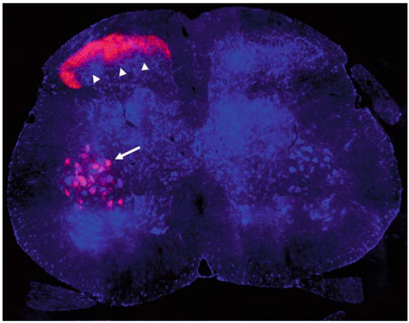
SPRR1A upregulation in the central process of primary afferent sensory neurons and in motoneurons after sciatic nerve injury. The sciatic nerve was crushed at the mid-thigh on one side of an adult mouse. Seven days later, the animal was sacrificed, and L5 spinal cord transverse sections were processed for anti-SPRR1A immunohistology (red) and for Nissl Stain (blue). Note the intense SPRR1A protein upregulation in afferent terminals in the dorsal horn (arrowheads) and in ventral horn motoneurons (arrow). Upregulation is confined to the injured side (left) with very low levels of SPRR1A on the intact side (right). Dorsal is up. Methods as in Bonilla et al. (2002). Image courtesy of Dr. William B. Cafferty
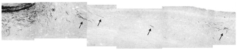
Corticospinal tract (CST) axonal tracing in mice lacking Nogo-A/B after mid-thoracic spinal cord dorsal hemisection. A parasagittal section of thoracic spinal cord from a mouse with a mutation in the Nogo gene that prevents Nogo-A and Nogo-B expression. The CST is traced from a cortical biotin dextran amine (BDA) injection after dorsal hemisection. Rostral is left; dorsal is up. The lesion is indicated by the asterisk. Note the evidence of CST fiber growth caudal to the lesion site (arrows). Significantly less BDA tracing is present caudal to the lesion in control animals (not shown). This photographic montage is a different image from the same mice described in Kim et al. (2003b)
References
-
- Atwal JK, Pinkston-Gosse J, Syken J, Stawicki S, Wu Y, Shatz C, Tessier-Lavigne M. PirB is a functional receptor for myelin inhibitors of axonal regeneration. Science. 2008;322:967–970. - PubMed
-
- Bartsch U, Bandtlow CE, Schnell L, Bartsch S, Spillmann AA, Rubin BP, Hillenbrand R, Montag D, Schwab ME, Schachner M. Lack of evidence that myelin-associated glycoprotein is a major inhibitor of axonal regeneration in the CNS. Neuron. 1995;15:1375–1381. - PubMed
-
- Benfey M, Aguayo AJ. Extensive elongation of axons from rat brain into peripheral nerve grafts. Nature. 1982;296:150–152. - PubMed
Publication types
MeSH terms
Substances
Grants and funding
LinkOut - more resources
Full Text Sources
Other Literature Sources
