

Percutaneous treatment of less severe intraarticular calcaneal fractures
- PMID: 19582524
- PMCID: PMC2835587
- DOI: 10.1007/s11999-009-0964-x
Percutaneous treatment of less severe intraarticular calcaneal fractures
Abstract
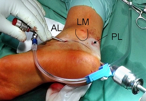
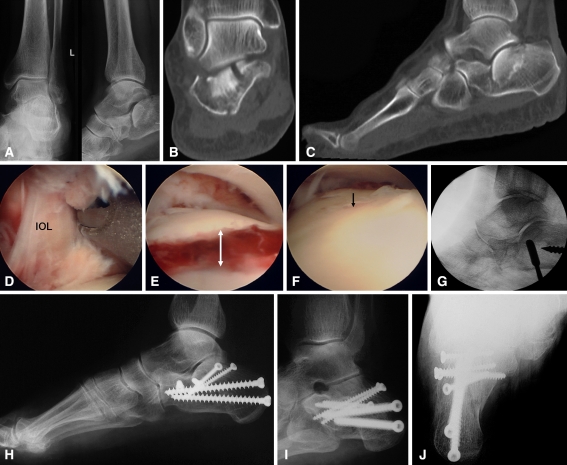
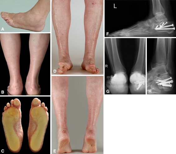
Percutaneous treatment of calcaneal fractures is intended to reduce soft tissue complications and postoperative stiffness of the subtalar joint. We assessed the complications, clinical hindfoot alignment, motion, functional outcome scores, and radiographic correction of percutaneous arthroscopically assisted reduction and screw fixation of selected, less severe fractures. We performed percutaneous reduction and screw fixation in 61 patients with Type II (Sanders et al.) calcaneal fractures. In 33 of 61 patients with displaced intraarticular fractures (types IIA and IIB), anatomic reduction of the subtalar joint was confirmed arthroscopically; these patients form the basis of this report. We observed no wound complications or infections. In two patients, one prominent screw was removed after 1 and 3 years, respectively. In one patient, arthroscopic arthrolysis was performed 1 year after the index procedure. Twenty-four of 33 patients (73%) were followed a minimum of 24 months (mean, 29 months; range, 24-67 months). The average American Orthopaedic Foot and Ankle Society ankle-hindfoot score at last followup was 92.1 (range, 80-100). Böhler's angle and calcaneal width were reduced close to the values of the uninjured side. We believe percutaneous fixation is a reasonable alternative for moderately displaced Type II fractures provided adequate control over anatomic joint reduction with either subtalar arthroscopy or high-resolution (3-D) fluoroscopy.
Level of evidence: Level IV, therapeutic study. See Guidelines for Authors for a complete description of levels of evidence.
Figures
References
-
- Abidi NA, Dhawan S, Gruen GS, Vogt MT, Conti SF. Wound-healing risk factors after open reduction and internal fixation of calcaneal fractures. Foot Ankle Int. 1998;19:856–861. - PubMed
-
- Benirschke SK, Sangeorzan BJ. Extensive intraarticular fractures of the foot: surgical management of calcaneal fractures. Clin Orthop Relat Res. 1993;292:128–134. - PubMed
-
- Bezes H, Massart P, Delvaux D, Fourquet JP, Tazi F. The operative treatment of intraarticular calcaneal fractures: indications, technique, and results in 257 cases. Clin Orthop Relat Res. 1993;290:55–59. - PubMed
-
- Böhler L. Behandlung der Fersenbeinbrüche. Arch Klin Chir. 1929;157:723–732. - PubMed
MeSH terms
LinkOut - more resources
Full Text Sources
Medical
