. 2009 Jul;39(7):1726-35.
doi: 10.1002/eji.200839001.

Multiple tissue-specific isoforms of sulfatide activate CD1d-restricted type II NKT cells

Affiliations

Multiple tissue-specific isoforms of sulfatide activate CD1d-restricted type II NKT cells

Maria Blomqvist et al. Eur J Immunol. 2009 Jul.

Abstract

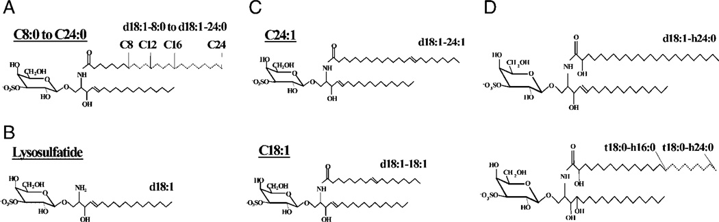
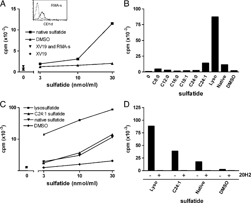
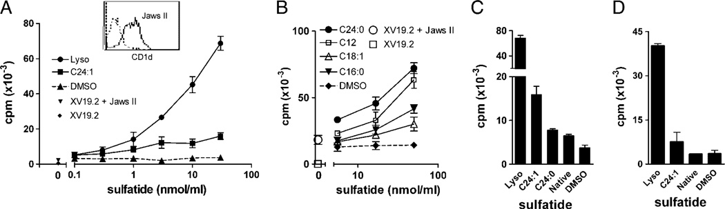
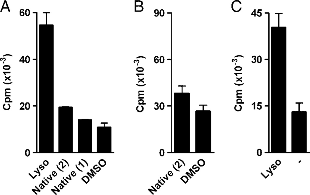
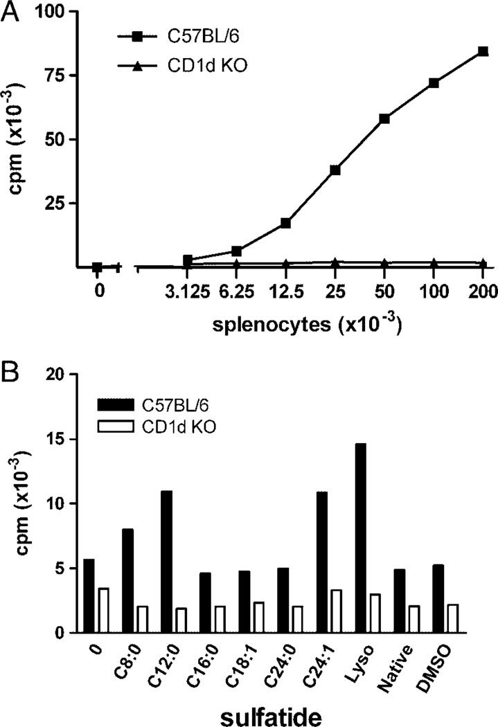
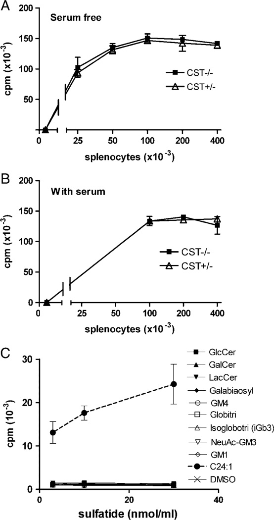
The glycosphingolipid sulfatide (SO(3)-3Galbeta1Cer) is a demonstrated ligand for a subset of CD1d-restricted NKT cells, which could regulate experimental autoimmune encephalomyelitis, a murine model for multiple sclerosis, as well as tumor immunity and experimental hepatitis. Native sulfatide is a mixture of sulfatide isoforms, i.e. sulfatide molecules with different long-chain bases and fatty acid chain lengths and saturation. Here, we demonstrate that sulfatide-specific CD1d-restricted murine NKT hybridomas recognized several different sulfatide isoforms. These included the physiologically relevant isoforms C24:1 and C24:0, major constituents of the myelin sheet of the nervous system, and C16:0, prominent in the pancreatic islet beta-cells. The most potent sulfatide isoform was lysosulfatide (lacking a fatty acid). Shortened fatty acid chain length (C24:1 versus C18:1), or saturation of the long fatty acid (C24:0), resulted in reduced stimulatory capacity, and fatty acid hydroxylation abolished the response. Moreover, sulfatide was not responsible for the natural autoreactivity toward splenocytes by XV19 T hybridoma cells. Our results reveal a promiscuity in the recognition of sulfatide isoforms by a CD1d-restricted NKT-cell clone, and suggest that sulfatide, a major component of the myelin sheet and pancreatic beta-cells, is one of several natural ligands for type II CD1d-restricted NKT cells.


Conflict of interest statement

Conflict of interest: The authors declare no financial or commercial conflict of interest.

Figures

Figure 1
Figure 1
Sulfatide isoforms. Semi-synthetic sulfatide isoforms with saturated fatty acids of C8–C24 length (A) and lyso (B) were synthesized from native sulfatide. Semi-synthetic C24:1 and C18:1 with unsaturated fatty acids are shown in (C). (D) Sulfatide isoforms were isolated from human intestine. The shorthand nomenclature for the sulfatide isoforms is indicated (see Table 1).
Figure 2
Figure 2
Activation of the NKT-cell hybridoma XV19 by distinct sulfatide isoforms using RMA-S cells as APC. RMA-S cells (50 × 103 cells/well) were pulsed with the indicated sulfatide isoforms or vehicle (DMSO) before the addition of XV19 NKT hybridoma cells (40 × 103 cells/well). Activation was measured as IL-2 secretion determined in a CTLL-2 bioassay as described in Materials and methods. (A) Different concentrations of native sulfatide (squares) or vehicle (DMSO) were used to stimulate XV19 NKT-cell hybridoma cells. Inset: CD1d expression of RMA-S cells was determined by flow cytometry (dotted line; unstained cells and solid line; CD1d–stained cells). (B) XV19 cells were stimulated with RMA-S cells pre-pulsed with 30 nmol/mL of the indicated sulfatide isoforms (see Fig. 1 and Table 1 for structures of sulfatide isoforms) or vehicle control. (C) XV19 cells were stimulated with a titration of selected sulfatide isoforms presented on RMA-S cells. (D) RMA-S cells were pre-pulsed with lysoe, C24:1 sulfatide or native sulfatide (30 nmol/mL), incubated with or without the CD1d mAb 20H2 [39] for 15 min, followed by addition of the XV19 NKT-cell hybridoma cells. Data shown are from one representative experiment of at least three (A–C) or two (D) (mean of duplicate cultures). Lyso, lysosulfatide.
Figure 3
Figure 3
The DC line JawsII efficiently presented multiple sulfatide isoforms to XV19 T hybridoma cells. JawsII cells (5 × 10³ cells per well) were incubated with the indicated sulfatides for 4h, followed by addition of XV19 NKT-cell hybridoma cells (40 × 103 cells per well). After 16 h, IL-2 secretion was assayed using CTLL-2 cells. Data shown are representative of three or more experiments (mean ± SD of triplicate cultures). (A) JawsII cells had been pre-pulsed with lyso or C24:1 at the indicated concentrations, before co-culture with XV19 NKT-cell hybridoma cells. Inset: CD1d expression of JawsII cells was determined by flow cytometry (dotted line; cells stained with isotype control antibody, and solid line; CD1d–stained cells). (B) JawsII cells were incubated together with titrations of indicated sulfatide isoforms, followed by the addition of XV19 NKT-cell hybridoma cells. (C) JawsII cells (5 × 10³) were cultured together with selected sulfatide isoforms or native sulfatide (30 nmol/mL) before stimulation of the XV19 NKT-cell hybridoma. (D) Bone marrow-derived DC (10 × 103 cells/well) were incubated with the indicated sulfatide preparations (30 nmol/mL) before stimulation of XV19 cells. Lyso, lysosulfatide.
Figure 4
Figure 4
Lyso strongly stimulated two different sulfatide reactive hybridomas. XV19 cells (A) or 14S.15.5D cells (B, C) were stimulated with APC pre-incubated with native sulfatide isolated from pig brain (Native (1), see Materials and methods) or commercially available native sulfatide from bovine brain (Native (2), Matreya), lyso, all at 30 nmol/ mL, or vehicle control (DMSO (A), or medium, –, (B)). XV19 cells were stimulated using JawsII (5 × 10³ cells/well), and 14S.15.5D with RAW-mCD1d cells (10 × 103 cells/well) as APC. Activation was measured in a CTLL-2 bioassay. Data are representative of at least three experiments performed (mean ± SD of triplicate cultures).
Figure 5
Figure 5
Spleen cells presented exogenous sulfatide to XV19 T hybridoma cells. (A) A titration of splenocytes was cultured together with the XV19 NKT-cell hybridoma, and IL-2 secretion assayed by CTLL-2 cells. Autoreactivity of the NKT-cell hybridoma XV19 was demonstrated to C57BL/6 (squares), but not CD1d−/− (CD1d KO, triangles) splenocytes. Data shown are representative of more than five experiments (mean of duplicate cultures). (B) Sulfatide pulsed CD1d+ splenocytes could stimulate XV19 cells above background autoreactivity. Splenocytes (12 × 103 cells/well) from C57BL/6 (black bars) or CD1d−/− (CD1d KO, white bars) mice were cultured together with sulfatide isoforms (30 nmol/mL) for 4 h before co-culture with the XV19 NKT-cell hybridoma (40 × 103 cells per well). Data shown are one representative experiment of three (mean of duplicate cultures). Lyso, lysosulfatide.
Figure 6
Figure 6
The NKT-cell hybridoma XV19 was autoreactive toward splenocytes lacking sulfatide. Indicated numbers of splenocytes from CST−/− (black squares) and CST+/− littermate control mice (white triangles) were cultured together with the XV19 NKT-cell hybridoma for 16 h, after which IL-2 secretion was assayed using CTLL-2 cells. The cultures were performed in the absence of serum (A) or in regular supplemented RPMI with 5% FBS (B). Data shown are one representative experiment of three (B) or one (A) performed (mean ± SD of triplicate cultures). (C) XV19 cells were tested for their reactivity to a number of different GSL (see Table 2) by pulsing JawsII cells (5 × 10³ cells/well) with the indicated GSL concentrations, before the addition of hybridoma cells. IL-2 secretion was assayed using CTLL-2 cells. Data are representative of two experiments using JawsII and two using RMA-S as APC.

References

    1. Bendelac A, Savage PB, Teyton L. The biology of NKT cells. Annu. Rev. Immunol. 2007;25:297–336. - PubMed
    1. Bricard G, Porcelli SA. Antigen presentation by CD1 molecules and the generation of lipid-specific T cell immunity. Cell. Mol. Life Sci. 2007;64:1824–1840. - PMC - PubMed
    1. Barral DC, Brenner MB. CD1 antigen presentation: how it works. Nat. Rev. Immunol. 2007;7:929–941. - PubMed
    1. Shamshiev A, Donda A, Carena I, Mori L, Kappos L, De Libero G. Self glycolipids as T-cell autoantigens. Eur. J. Immunol. 1999;29:1667–1675. - PubMed
    1. Shamshiev A, Gober HJ, Donda A, Mazorra Z, Mori L, De Libero G. Presentation of the same glycolipid by different CD1 molecules. J. Exp. Med. 2002;195:1013–1021. - PMC - PubMed

Publication types

MeSH terms

LinkOut - more resources