. 2009 Oct 15;125(8):1965-74.
doi: 10.1002/ijc.24530.

Oncogenic microRNA-27a is a target for anticancer agent methyl 2-cyano-3,11-dioxo-18beta-olean-1,12-dien-30-oate in colon cancer cells

Affiliations

Oncogenic microRNA-27a is a target for anticancer agent methyl 2-cyano-3,11-dioxo-18beta-olean-1,12-dien-30-oate in colon cancer cells

Sudhakar Chintharlapalli et al. Int J Cancer. .

Abstract

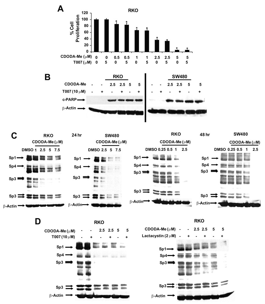
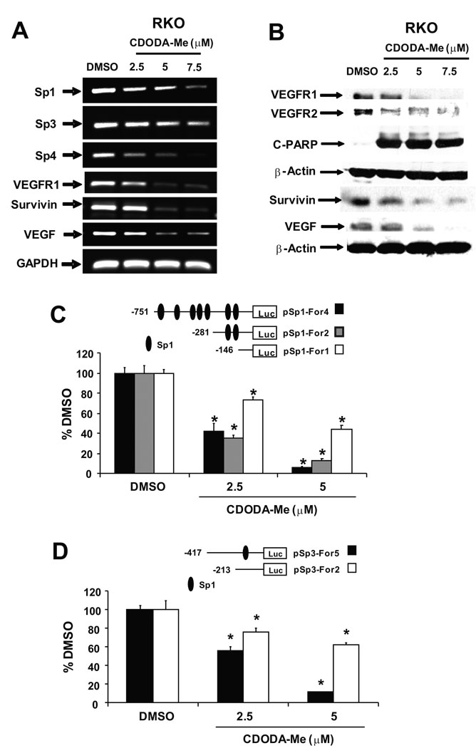
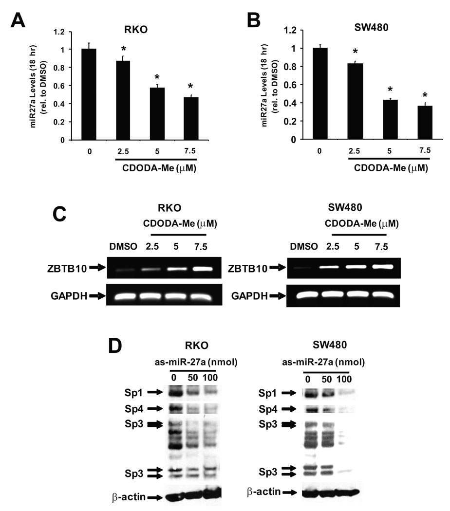
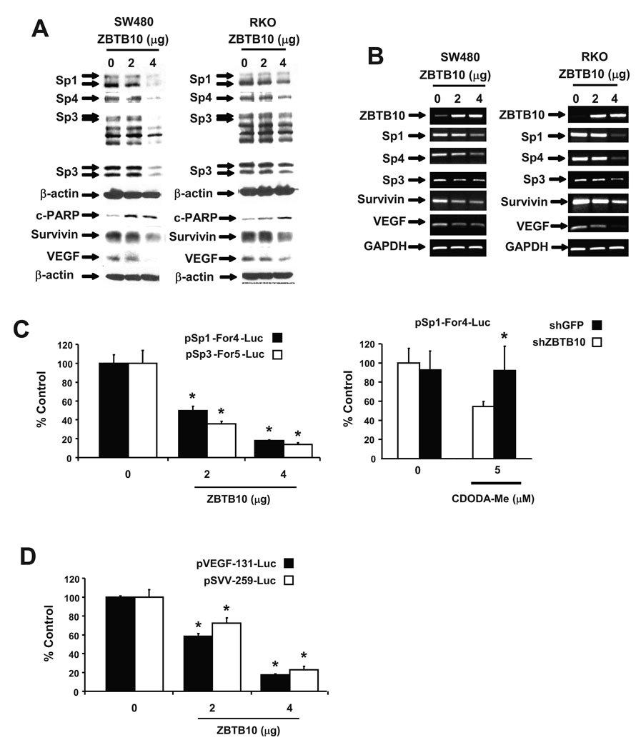
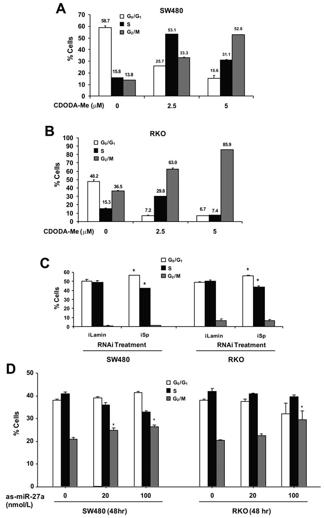
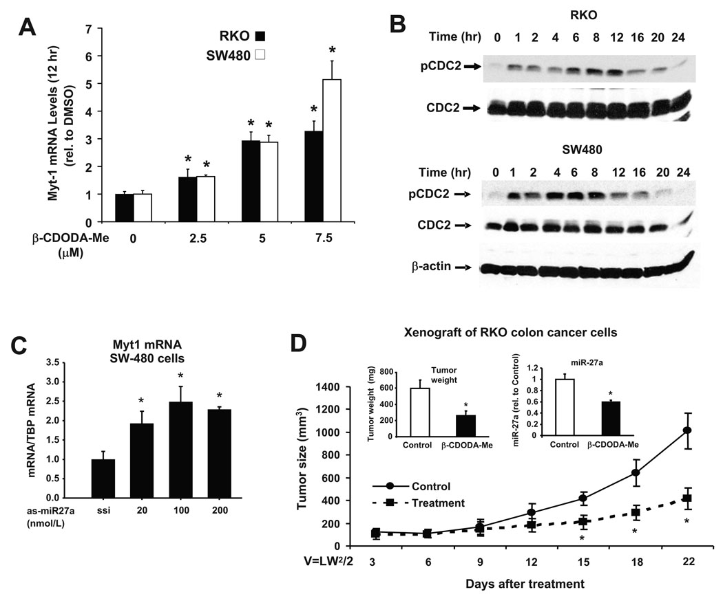
Methyl 2-cyano-3,11-dioxo-18beta-olean-1,12-dien-30-oate (CDODA-Me) is a synthetic derivative of glycyrrhetinic acid, a triterpenoid phytochemical found in licorice extracts. CDODA-Me inhibited growth of RKO and SW480 colon cancer cells and this was accompanied by decreased expression of Sp1, Sp3 and Sp4 protein and mRNA and several Sp-dependent genes including survivin, vascular endothelial growth factor (VEGF), and VEGF receptor 1 (VEGFR1 or Flt-1). CDODA-Me also induced apoptosis, arrested RKO and SW480 cells at G(2)/M, and inhibited tumor growth in athymic nude mice bearing RKO cells as xenografts. CDODA-Me decreased expression of microRNA-27a (miR-27a), and this was accompanied by increased expression of 2 miR-27a-regulated mRNAs, namely ZBTB10 (an Sp repressor) and Myt-1 which catalyzes phosphorylation of cdc2 to inhibit progression of cells through G(2)/M. Both CDODA-Me and antisense miR-27a induced comparable responses in RKO and SW480 cells, suggesting that the potent anticarcinogenic activity of CDODA-Me is due to repression of oncogenic miR-27a.


Conflict of interest statement

Conflict of Interest Statement: None declared.

Figures

Figure 1
Figure 1
CDODA-Me inhibits growth, induces cleaved PARP, and degradation of Sp proteins. (A) Decreased cell proliferation in RKO cells. Cells were seeded and treated with solvent (DMSO) or different concentrations of CDODA-Me (0.5–5 µM) alone or in combination with T007 for 4 days. Cell proliferation is expressed as the percentage of CDODA-Me-treated cells remaining compared to DMSO (set at 100%). Results are expressed as means ± SE for three replicate determinations for each treatment group and significantly (p < 0.05) decreased proliferation is indicated by an asterisk. Similar results were observed for SW480 cells (data not shown). Induction of cleaved PARP (B) and decreased expression of Sp1, Sp3 and Sp4 (C, D). RKO and SW480 cells were treated with DMSO, CDODA-Me (1–5 µM), T007 (10 µM), Lactacystin (2 µM), or combinations as indicated for 24 hr or 48 hr and whole cell lysates were analyzed by Western blot analysis as described in the Materials and Methods.
Figure 2
Figure 2
Effects of CDODA-Me on Sp and Sp-dependent expression. CDODA-Me decreases expression of Sp1, Sp3 and Sp4 and Sp-dependent angiogenic/survival gene mRNAs (A) and proteins (B). RKO cells were treated with different concentrations of CDODA-Me and after 24 hr, mRNA and protein were extracted and analyzed by semi-quantitative RT-PCR and Western blots, respectively, as described in the Materials and Methods. CDODA-Me decreases Sp1 (C) and Sp3 (D) promoter activity. RKO cells were transfected with various constructs, treated with different concentrations of CDODA-Me, and luciferase activity determined as described in the Materials and Methods. Results are expressed as means ± SE for three replicate determinations for each treatment group and significantly (p < 0.05) decreased activity is indicated by an asterisk.
Figure 3
Figure 3
Effects of CDODA-Me and as-miR27a. CDODA-Me decreases miR-27a (A and B) and increases ZBTB10 (C) expression. RKO and SW480 colon cancer cells were treated with DMSO or different concentrations of CDODA-Me and after 18 hr, total RNA was extracted and analyzed for miR27a by real time PCR and ZBTB10 by semi-quantitative RT-PCR as described in the Materials and Methods. Northern blot analysis also showed that CDODA-Me decreased miR-27a in colon cancer cells (data not shown). (D) as-miR27a decreases Sp1, Sp4 and Sp3 proteins. RKO cells were transfected with 50 and 100 ng as-miR27a and after 24 hr, whole cell lysates were analyzed by Western blot analysis for Sp1, Sp4 and Sp3 proteins as described in the Materials and Methods.
Figure 4
Figure 4
ZBTB10 decreases expression of Sp proteins and Sp-dependent angiogenic and survival genes. ZBTB10 expression decreases expression of Sp and angiogenic/survival proteins (A) and mRNA (B). RKO and SW480 cells were transfected with ZBTB10 expression plasmid and after 24 hr, protein and mRNA were extracted and analyzed by Western blots and semi-quantitative RT-PCR, respectively, as described in the Materials and Methods. (C) Role of ZBTB10 in mediating CDODA-Me-dependent downregulation of Sp1 and Sp3 promoter activity. RKO cells were transfected with pSp1-For4-luc or pSp3-For5-luc and cotransfected with ZBTB10 (left) or cotransfected with pSp1-For4-luc and shGFP or shZBTB10 and treated with DMSO or CDODA-Me (right), and luciferase activity was determined as described in the Materials and Methods. (D) ZBTB10 expression decreases VEGF and survivin promoter activity. RKO cells were transfected with various constructs and ZBTB10 expression plasmid, and luciferase activity was determined as described in the Materials and Methods. Results in (C) and (D) are expressed as means ± SE for three replicate determinations for each treatment group and significantly (p < 0.05) decreased activity is indicated by an asterisk.
Figure 5
Figure 5
Modulation of cell cycle progression by CDODA-Me, RNA interference and as-miR-27a. Effects of CDODA-Me. SW480 (A) and RKO (B) cells were treated for 24 hr with DMSO (0), 2.5 and 5.0 µM CDODA-Me, and analyzed by FACS analysis as described in the Materials and Methods. (C) iSp modulates Sp protein expression and the cell cycle in SW480 and RKO cells. Cells were transfected with iSp, a combination of small inhibitory RNAs for Sp1, Sp3 and Sp4 or a non-specific oligonucleotide (iLamin), and analyzed for Sp proteins by Western blots (to confirm Sp knockdown) (see Supplemental data) and FACS analysis as described in the Materials and Methods. (D) as-miR-27a modulates cell cycle progression. SW480 and RKO cells were transfected with different amounts of as-miR-27a and, after 48 hr, analyzed by FACS as described in the Materials and Methods. All experiments in this Figure [(A) – (D)] were repeated three times, and results are expressed as means ± SE. Significant (p < 0.05) changes compared to untreated (0) or iLamin-treated cells are indicated by asterisks.
Figure 6
Figure 6
In vitro and in vivo effects of CDODA-Me on G2/M arrest and inhibition of tumor growth. Effects of CDODA-Me (A and B) and as-miR-27a (C) on Myt-1 and cdc2 phosphorylation. Colon cancer cells were treated with different amounts of CDODA-Me or as-miR-27a for the indicated times, and Myt-1 expression and cdc2 phosphorylation were determined by real time PCR or Western blots, respectively, as described in the Materials and Methods. The effects of as-miR-27a on Myt-1 mRNA expression in RKO cells was similar to that observed in SW480 cells, and as-miR-27a also increased cdc2 expression as described (7) (data not shown). (D) CDODA-Me inhibits tumor growth (volume) and weight/miR-27a expression in a mouse xenograft model. Athymic nude mice bearing RKO cells as xenografts were treated with corn oil (solvent control) or CDODA-Me (15 mg/kg/d), and tumor volumes, tumor weights, and miR-27a expression were determined as described in the Materials and Methods. Results are expressed as means ± SE for replicate (at least three or more) determinations for each treatment group, and significantly (p < 0.05) decreased tumor volume or weight and miR-27a is indicated by an asterisk.

References

    1. Lewis BP, Burge CB, Bartel DP. Conserved seed pairing, often flanked by adenosines, indicates that thousands of human genes are microRNA targets. Cell. 2005;120:15–20. - PubMed
    1. Krek A, Grun D, Poy MN, Wolf R, Rosenberg L, Epstein EJ, MacMenamin P, da PI, Gunsalus KC, Stoffel M, Rajewsky N. Combinatorial microRNA target predictions. Nat Genet. 2005;37:495–500. - PubMed
    1. Zhu S, Si ML, Wu H, Mo YY. MicroRNA-21 targets the tumor suppressor gene tropomyosin 1 (TPM1) J Biol Chem. 2007;282:14328–14336. - PubMed
    1. Johnson SM, Grosshans H, Shingara J, Byrom M, Jarvis R, Cheng A, Labourier E, Reinert KL, Brown D, Slack FJ. RAS is regulated by the let-7 microRNA family. Cell. 2005;120:635–647. - PubMed
    1. Lee YS, Dutta A. The tumor suppressor microRNA let-7 represses the HMGA2 oncogene. Genes Dev. 2007;21:1025–1030. - PMC - PubMed

Publication types

MeSH terms