. 2009 Jul;10(7):883-93.
doi: 10.1111/j.1600-0854.2009.00915.x.

PIKfyve regulation of endosome-linked pathways

Affiliations
Free PMC article

PIKfyve regulation of endosome-linked pathways

Jane de Lartigue et al. Traffic. 2009 Jul.
Free PMC article

Abstract

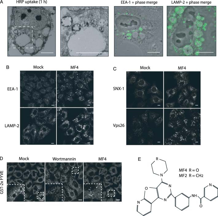
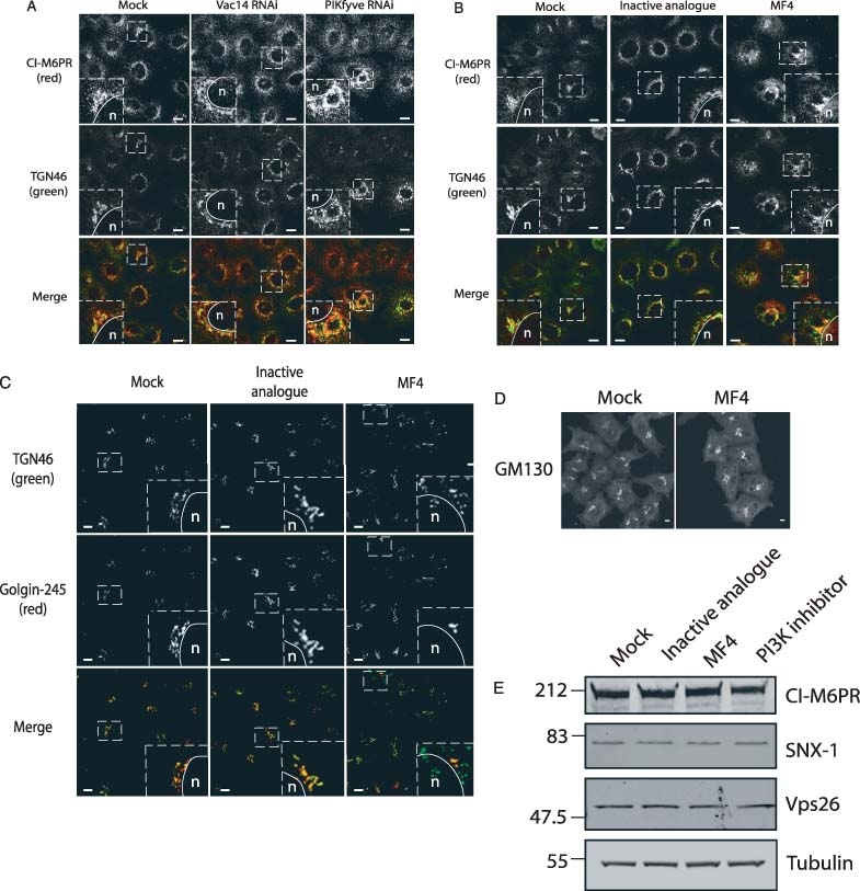
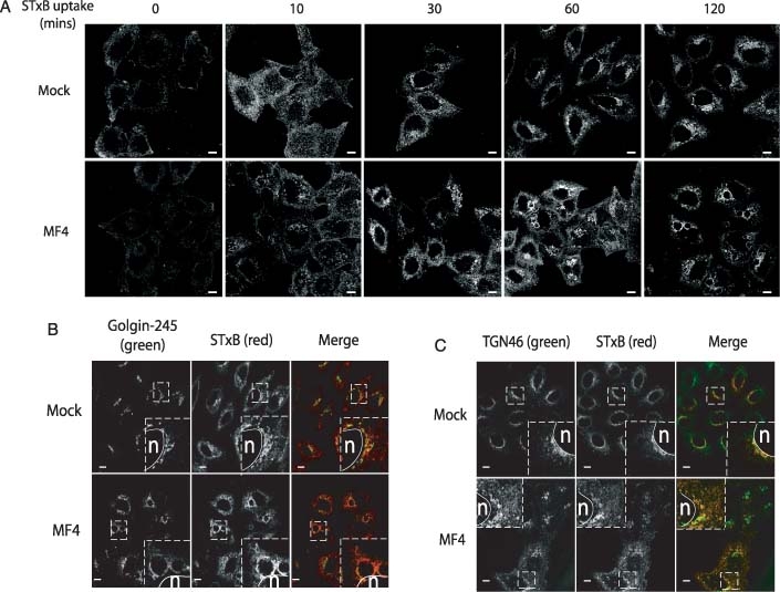
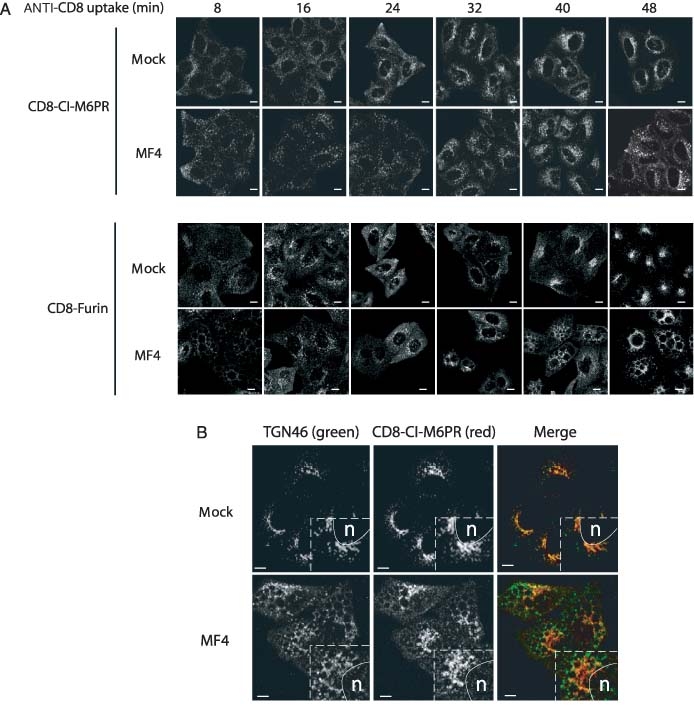
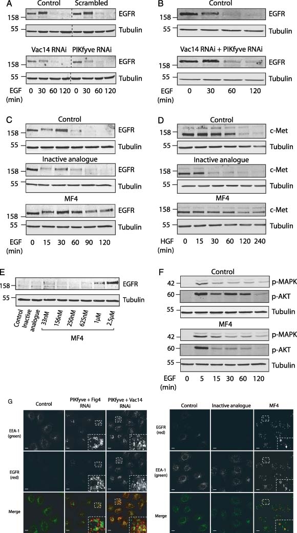
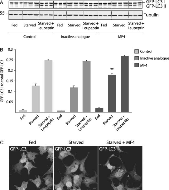
The phosphoinositide 5-kinase (PIKfyve) is a critical enzyme for the synthesis of PtdIns(3,5)P2, that has been implicated in various trafficking events associated with the endocytic pathway. We have now directly compared the effects of siRNA-mediated knockdown of PIKfyve in HeLa cells with a specific pharmacological inhibitor of enzyme activity. Both approaches induce changes in the distribution of CI-M6PR and trans-Golgi network (TGN)-46 proteins, which cycles between endosomes and TGN, leading to their accumulation in dispersed punctae, whilst the TGN marker golgin-245 retains a perinuclear disposition. Trafficking of CD8-CI-M6PR (retromer-dependent) and CD8-Furin (retromer-independent) chimeras from the cell surface to the TGN is delayed following drug administration, as is the transport of the Shiga toxin B-subunit. siRNA knockdown of PIKfyve produced no defect in epidermal growth factor receptor (EGFR) degradation, unless combined with knockdown of its activator molecule Vac14, suggesting that a low threshold of PtdIns(3,5)P2 is necessary and sufficient for this pathway. Accordingly pharmacological inhibition of PIKfyve results in a profound block to the lysosomal degradation of activated epidermal growth factor (EGF) and Met receptors. Immunofluorescence revealed EGF receptors to be trapped in the interior of a swollen endosomal compartment. In cells starved of amino acids, PIKfyve inhibition leads to the accumulation of the lipidated form of GFP-LC3, a marker of autophagosomal structures, which can be visualized as fluorescent punctae. We suggest that PIKfyve inhibition may render the late endosome/lysosome compartment refractory to fusion with both autophagosomes and with EGFR-containing multivesicular bodies.


Figures

Figure 1
Figure 1. PIKfyve inhibition creates large swollen vacuoles inaccessible to fluid phase marker
HeLa cells were treated with 800 nm PIKfyve inhibitor (MF4) for 4 h prior to either fixation and staining with a variety of markers by immunofluorescence, or uptake of HRP for 1 h, fixation and processing for EM (see Materials and Methods for details). Wortmannin was added to cells at 100 nm for 30 min. A) MF4-mediated inhibition of PIKfyve causes the formation of swollen vacuoles that are inaccessible to internalized HRP. These vacuoles are surrounded by a second class of smaller HRP-containing vacuoles. B) Both early and late endosomal markers are associated with distinct swollen endosomes that surround these vacuoles. C) Retromer components Vps26 and SNX-1 also associate with swollen endosomal compartments. Scale bars = 10 μm. D) The PtdIns 3-kinase inhibitor Wortmannin ablates endosomal PtdIns(3)P staining, whilst MF4 treatment has no significant effect. Scale bars = 20 μm. E) Chemical structure of MF4 (PIKfyve inhibitor) and MF2 (inactive analogue).
Figure 2
Figure 2. Inhibition of PIKfyve causes a disruption in steady-state distribution of ciM6PR and TGN46
HeLa cells were treated with 40 nM siRNA oligos for 72 h or 800 nm MF4 for 4 h. Both knockdown and inhibition of PIKfyve alter the steady-state localization of ciM6PR, whereas Vac14 knockdown and MF2 (inactive analogue of the PIKfyve inhibitor MF4) have no effect (A and B). The markers fragment into distinct vesicular compartments, which show little co-localization. The distribution of a second TGN resident protein golgin-245 is unaffected and retains its characteristic perinuclear localization (D). The cis-golgi marker GM130 also retains it's ribbon-like disposition (E). There is no change in the levels of ciM6PR or retromer components upon treatment with PIKfyve inhibitor (F). Scale bars = 20 μm.
Figure 4
Figure 4. PIKfyve inhibition causes a perturbation in the uptake of the Shiga toxin B-subunit
Uptake of the Shiga toxin B-subunit was examined in HeLa cells using a Cy3-labelled StxB construct, following treatment with 800 nm MF4 PIKfyve inhibitor. Cy3-STxB retrieval to a perinuclear localization is also delayed in cells treated with PIKfyve inhibitor (A), but ultimately reaches a TGN localization, as judged by co-localization with golgin-245 (B). In cells where TGN46 is highly dispersed, co-localization with Cy3-STxB can be observed, indicative of the fact that these two proteins may traffic on a similar retrieval pathway (C). Scale bars = 20 μm.
Figure 3
Figure 3. PIKfyve inhibition causes a delay in CD8-ciM6PR and CD8-Furin retrieval
Uptake of CD8 antibody in HeLa M cell lines stably expressing CD8-ciM6PR and CD8-Furin was examined following treatment of cells with 800 nm MF4 PIKfyve inhibitor. Both Furin and ciM6PR retrieval to a trans-Golgi localization is delayed upon inhibition of PIKfyve (A). At the terminal uptake time-point, in cells where TGN46 is highly fragmented, there is negligible co-localization with CD8-ciM6PR (B). Scale bars = 20 μm.
Figure 5
Figure 5. Acute inhibition of PIKfyve affects downregulation of tyrosine kinase receptors
HeLa cells were treated with siRNA oligos against the indicated proteins (A and B), or with 3 μm PIKfyve inhibitor (C and D) or the indicated doses of inhibitor (E). Serum-starved cells were stimulated with 100 ng/mL EGF or HGF for the indicated times. A) Individual knockdown of PIKfyve or Vac14 did not affect the degradation of either EGFR or c-Met. B) A combined knockdown of PIKfyve with Vac14 caused a modest delay in receptor downregulation. C) Treatment with PIKfyve inhibitor caused a severe delay in EGFR. D) c-Met downregulation. E) Concentration dependence of MF4 on EGFR downregulation after 120 min EGF stimulation. F) pMAPK and pAKT signalling is not prolonged following treatment with PIKfyve inhibitor, pAKT signalling is dampened at later time-points. α-tubulin is used as a protein loading control. G) Immunofluorescence images demonstrating the accumulation of EGFR in PIKfyve inhibitor and combined knockdown treated HeLa cells. HeLa cells were transfected with 40 nM of the indicated oligos over 72 h, then serum-starved overnight, and following treatment with 800 nm PIKfyve inhibitor (in place of siRNA treatment; where indicated), were stimulated with 100 ng/mL EGF for 4 h. Following fixation with 3% PFA in PBS cells were labelled with anti-EGFR and anti-EEA-1. A proportion of the EGFR can be seen in the interior of swollen early endosomal compartments (G). Scale bars = 20 μm.
Figure 6
Figure 6. Inhibition of PIKfyve leads to an increase in starvation-induced GFP-LC3 lipidation
HEK293A cells stably transfected with GFP-LC3 were treated with MF4 PIKfyve inhibitor or an inactive analogue (MF2) for 4 h prior to replacement of fresh growth medium (fed) or Earles’ balanced salt solution (EBSS) for 2 h (starved). Cells were subsequently lysed or processed for immunofluorescence. A) Western blot on triplicate samples indicating the levels of GFP-LC3 I and II in fed and starved cells upon treatment with MF4 and with or without the protease inhibitor Leupeptin. B) Quantitative analysis of the ratio of GFP-LC3 II to total GFP-LC3 from A. PIKfyve inhibition causes a statistically significant (n = 3, p < 0.01, T-test) accumulation of lipidated GFP-LC3 upon starvation-induced autophagy. C) Immunofluorescence analysis of GFP-LC3 II punctae formation following starvation-induced autophagy. The accumulation of GFP-LC3 II positive punctae can be observed following treatment with PIKfyve inhibitor. Scale bars = 20 μm.

References

    1. Lindmo K, Stenmark H. Regulation of membrane traffic by phosphoinositide 3-kinases. J Cell Sci. 2006;119:605–614. - PubMed
    1. Joly M, Kazlauskas A, Corvera S. Phosphatidylinositol 3-kinase activity is required at a postendocytic step in platelet-derived growth-factor receptor trafficking. J Biol Chem. 1995;270:13225–13230. - PubMed
    1. Jones AT, Clague MJ. Phosphatidylinositol 3-kinase activity is required for early endosome fusion. Biochem J. 1995;311:31–34. - PMC - PubMed
    1. Mills IG, Jones AT, Clague MJ. Involvement of the endosomal autoantigen EEA1 in homotypic fusion of early endosomes. Curr Biol. 1998;8:881–884. - PubMed
    1. Raiborg C, Bache KG, Gillooly DJ, Madshus IH, Stang E, Stenmark H. Hrs sorts ubiquitinated proteins into clathrin-coated microdomains of early endosomes. Nat Cell Biol. 2002;4:394–398. - PubMed

Publication types

MeSH terms

Substances