Review
. 2009 Dec;15(4):477-84.
doi: 10.1089/ten.TEB.2009.0340.

The impact of biomechanics in tissue engineering and regenerative medicine

Affiliations
Review

The impact of biomechanics in tissue engineering and regenerative medicine

David L Butler et al. Tissue Eng Part B Rev. 2009 Dec.

Abstract

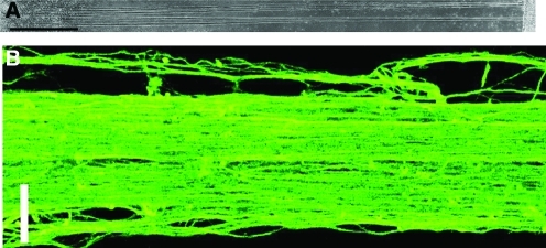
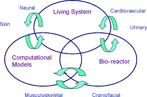
Biomechanical factors profoundly influence the processes of tissue growth, development, maintenance, degeneration, and repair. Regenerative strategies to restore damaged or diseased tissues in vivo and create living tissue replacements in vitro have recently begun to harness advances in understanding of how cells and tissues sense and adapt to their mechanical environment. It is clear that biomechanical considerations will be fundamental to the successful development of clinical therapies based on principles of tissue engineering and regenerative medicine for a broad range of musculoskeletal, cardiovascular, craniofacial, skin, urinary, and neural tissues. Biomechanical stimuli may in fact hold the key to producing regenerated tissues with high strength and endurance. However, many challenges remain, particularly for tissues that function within complex and demanding mechanical environments in vivo. This paper reviews the present role and potential impact of experimental and computational biomechanics in engineering functional tissues using several illustrative examples of past successes and future grand challenges.


Figures

FIG. 1.
FIG. 1.
Effects of dynamic loading on the mechanical properties of cartilage contstructs. Mechanical preconditioning was carried out with three intermittent (separated by 1 h) loading cycles per day of 10% deformation at 1 Hz. Loading was then carried out for 5 days per week for a total of 4 weeks. *, Indicates significant differences between loaded samples and free-swelling controls; **, indicates a significant difference between peak stress of day 21 and day 28 loaded samples.
FIG. 2.
FIG. 2.
Histology of engineered vessels. Cultured for 8 weeks revealed by Masson's Trichrome stain (collagen stains blue). (B) Cyclically stretched. (D) Nonstretched (original magnification × 100). Number sign indicates the dense cellular region; asterisk indicates the residual polymer region. Reproduced with permission from Niklason et al. Color images available online at www.liebertonline.com/ten.
FIG. 3.
FIG. 3.
(A) Three-dimensional Lattice–Boltzmann simulation of local shear stresses resulting from fluid flow through a porous polycaprolactone scaffold (white) seeded with marrow-derived progenitor cells in a perfusion bioreactor. (B) Micro-CT images of mineralized matrix synthesis in perfused constructs compared with constructs cultured under static conditions. (C) 9 mm long mineralized construct produced under dynamic culture using stem cells seeded on a polycaprolactone scaffold. Color images available online at www.liebertonline.com/ten.
FIG. 4.
FIG. 4.
Fiber alignment in (a) native leaflet, (b) 3 week valve-equivalent leaflet, and (c) 3 week valve-equivalent root as determined by polarized light imaging. The orientations of the white lines correspond to the local average fiber direction, and their lengths are proportional to the local average retardation, a measure of the fiber alignment strength. Simulation of (d) gel compaction in the mold, and (e) principal stresses based on the anisotropic biphasic theory of tissue-equivalent mechanics. Black lines indicate principal fiber alignment directions in the root (triangulated gray area) and leaflet (solid white and gray interior areas) in (d) and directions of principal stresses in (e). Reproduced with permission from Robinson et al. Color images available online at www.liebertonline.com/ten.
FIG. 5.
FIG. 5.
Representative images of axon tracts after 7 days of stretch induced growth. (A) Phase micrograph showing tracts between neurons adherent on membranes at both ends (scale bar = 1 mm). (B) Confocal micrograph of immunostained microtubule protein of coalescing axons in a single tract (scale bar = 25 μm). Reproduced with permission from Smith et al. Color images available online at www.liebertonline.com/ten.
FIG. 6.
FIG. 6.
Schematic illustrating the interactions required among in vivo living system, in vitro bioreactor, and computational models necessary to understand the influence of biomechanics on tissue repair and regeneration. Representative applications that will benefit from research in these strategic areas are included around the Venn diagram. Color images available online at www.liebertonline.com/ten.

References

    1. Carrel A. Lindberg C. The Culture of Organs. The Canadian Medical Association Journal. New York: Paul B. Hoeber, Inc., Medical Book Department of Harper & Brothers; 1938.
    1. Skalak R. Fox C. NSF Workshop: UCLA Symposia on Molecular and Cellular Biology. Molecular and Cellular Biology, New Series. New York: Alan R. Liss, Inc.; 1988.
    1. Butler D.L. Goldstein S.A. Guilak F. Functional tissue engineering: the role of biomechanics. J Biomech Eng. 2000;122:570. - PubMed
    1. Engler A.J. Rehfeldt F. Sen S. Discher D.E. Microtissue elasticity: measurements by atomic force microscopy and its influence on cell differentiation. Methods Cell Biol. 2007;83:521. - PubMed
    1. Engler A.J. Sen S. Sweeney H.L. Discher D.E. Matrix elasticity directs stem cell lineage specification. Cell. 2006;126:677. - PubMed