Clinical Trial
. 2009 Sep;20(9):999-1004.
doi: 10.1089/hum.2009.086.

Human RPE65 gene therapy for Leber congenital amaurosis: persistence of early visual improvements and safety at 1 year

Affiliations
Clinical Trial

Human RPE65 gene therapy for Leber congenital amaurosis: persistence of early visual improvements and safety at 1 year

Artur V Cideciyan et al. Hum Gene Ther. 2009 Sep.

Abstract

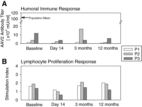
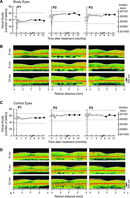
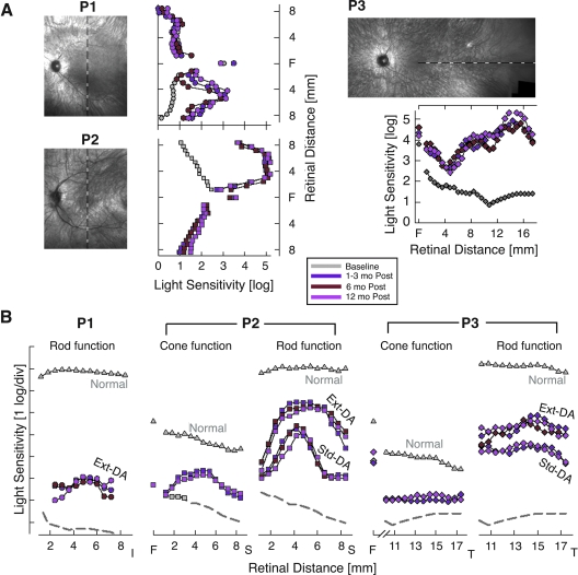
Human gene therapy with rAAV2-vector was performed for the RPE65 form of childhood blindness called Leber congenital amaurosis. In three contemporaneous studies by independent groups, the procedure was deemed safe and there was evidence of visual gain in the short term. At 12 months after treatment, our young adult subjects remained healthy and without vector-related serious adverse events. Results of immunological assays to identify reaction to AAV serotype 2 capsid were unchanged from baseline measurements. Results of clinical eye examinations of study and control eyes, including visual acuities and central retinal structure by in vivo microscopy, were not different from those at the 3-month time point. The remarkable improvements in visual sensitivity we reported by 3 months were unchanged at 12 months. The retinal extent and magnitude of rod and cone components of the visual sensitivity between 3 and 12 months were also the same. The safety and efficacy of human retinal gene transfer with rAAV2-RPE65 vector extends to at least 1 year posttreatment.


Figures

FIG. 1.
FIG. 1.
Immunological assays before surgery (baseline) and at 14-, 90-, and 365-day time points after retinal gene therapy. (A) Humoral immune response to AAV serotype 2 (AAV2) assayed by circulating serum antibody titers against AAV2 capsid in P1, P2, and P3 before and after surgery. Arrow along the vertical axis indicates mean levels from a normal reference population (n = 79) (Hauswirth et al., 2008). (B) Antigen-specific lymphocyte proliferation response (ASR) assayed in peripheral blood lymphocytes incubated in the presence versus in the absence of AAV2 capsid antigen. The stimulation index (the ratio of [3H]thymidine uptake in the presence of antigen to the uptake in its absence) in each subject after surgery is unchanged from baseline values.
FIG. 2.
FIG. 2.
Central vision and retinal structure in RPE65-LCA during the 12 months after gene therapy. (A) Visual acuity as a function of time before and after the day of surgery (0) in the study eyes of P1, P2, and P3. Gray symbols, data previously reported (Hauswirth et al., 2008). Gray lines, 15-letter gain or loss from baseline visual acuity. (B) Cross-sectional optical coherence tomography (OCT) scans of retina along the horizontal meridian 6, 9, and 12 months after surgery. Overlaid white lines represent the location of the vitreoretinal boundary 3 months posttreatment (Hauswirth et al., 2008). (C) Visual acuity and (D) OCT scans in untreated control eyes for comparison. F, fovea; N, nasal; T, temporal retina.
FIG. 3.
FIG. 3.
Visual function improvement due to gene therapy is stable up to 12 months after treatment. (A) Light sensitivity to achromatic stimuli in study eyes along vertical (P1 and P2) and horizontal (P3) meridians after allowing for an extended (3–8 hr) period of dark adaptation. Sensitivities 6 and 12 months after treatment are compared with the mean value at 1, 2, and 3 months after treatment or the mean baseline value before treatment. Loci of visual function testing are shown on images of the ocular fundus of the study eyes obtained at 12 months. All images are shown as left retina for clarity and comparability. F, fovea. (B) Light sensitivity measures with chromatic stimuli support stability of rod and cone photoreceptor-based vision 12 months after treatment in retinal regions of peak response to gene therapy. Rod function measured with blue stimuli after standard (Std) or extended dark adaptation (Ext-DA) conditions. Cone function measured with red stimuli after Ext-DA conditions (at the fovea) or during the cone plateau period in the dark after light adaptation (at extrafoveal locations). The sensitivity axes are shifted vertically to match red and blue stimuli for cone-mediated detection in patients. I, inferior; S, superior; T, temporal retina.

References

    1. Acland G.M. Aguirre G.D. Bennett J. Aleman T.S. Cideciyan A.V. Bennicelli J. Dejneka N.S. Pearce-Kelling S.E. Maguire A.M. Palczewski K. Hauswirth W.W. Jacobson S.G. Long-term restoration of rod and cone vision by single dose rAAV-mediated gene transfer to the retina in a canine model of childhood blindness. Mol. Ther. 2005;12:1072–1082. - PMC - PubMed
    1. Aguirre G.K. Komáromy A.M. Cideciyan A.V. Brainard D.H. Aleman T.S. Roman A.J. Avants B.B. Gee J.C. Korczykowski M. Hauswirth W.W. Acland G.M. Aguirre G.D. Jacobson S.G. Canine and human visual cortex intact and responsive despite early retinal blindness from RPE65 mutation. PLoS Med. 2007;4:e230. - PMC - PubMed
    1. Aleman T.S. Cideciyan A.V. Sumaroka A. Windsor E.A. Herrera W. White D.A. Kaushal S. Naidu A. Roman A.J. Schwartz S.B. Stone E.M. Jacobson S.G. Retinal laminar architecture in human retinitis pigmentosa caused by rhodopsin gene mutations. Invest. Ophthalmol. Vis. Sci. 2008;49:1580–1590. - PMC - PubMed
    1. Bainbridge J.W. Smith A.J. Barker S.S. Robbie S. Henderson R. Balaggan K. Viswanathan A. Holder G.E. Stockman A. Tyler N. Petersen-Jones S. Bhattacharya S.S. Thrasher A.J. Fitzke F.W. Carter B.J. Rubin G.S. Moore A.T. Ali R.R. Effect of gene therapy on visual function in Leber's congenital amaurosis. N. Engl. J. Med. 2008;358:2231–2239. - PubMed
    1. Cai X. Conley S.M. Naash M.I. RPE65: Role in the visual cycle, human retinal disease, and gene therapy. Ophthalmic Genet. 2009;30:57–62. - PMC - PubMed

Publication types

MeSH terms