Review
. 2009 Sep;13(9B):2990-3005.
doi: 10.1111/j.1582-4934.2009.00831.x. Epub 2009 Jul 6.

Histone deacetylase inhibition and the regulation of cell growth with particular reference to liver pathobiology

Affiliations
Review

Histone deacetylase inhibition and the regulation of cell growth with particular reference to liver pathobiology

Fraczek Joanna et al. J Cell Mol Med. 2009 Sep.

Abstract

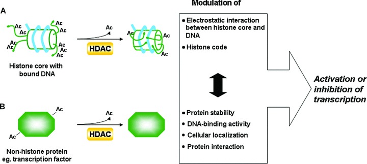
The transcriptional activity of genes largely depends on the accessibility of specific chromatin regions to transcriptional regulators. This process is controlled by diverse post-transcriptional modifications of the histone amino termini of which reversible acetylation plays a vital role. Histone acetyltransferases (HATs) are responsible for the addition of acetyl groups and histone deacetylases (HDACs) catalyse the reverse reaction. In general, though not exclusively, histone acetylation is associated with a positive regulation of transcription, whereas histone deacetylation is correlated with transcriptional silencing. The elucidation of unequivocal links between aberrant action of HDACs and tumorigenesis lies at the base of key scientific importance of these enzymes. In particular, the potential benefit of HDAC inhibition has been confirmed in various tumour cell lines, demonstrating antiproliferative, differentiating and pro-apoptotic effects. Consequently, the dynamic quest for HDAC inhibitors (HDIs) as a new class of anticancer drugs was set off, resulting in a number of compounds that are currently evaluated in clinical trials. Ironically, the knowledge with respect to the expression pattern and function of individual HDAC isoenzymes remains largely elusive. In the present review, we provide an update of the current knowledge on the involvement of HDACs in the regulation of fundamental cellular processes in the liver, being the main site for drug metabolism within the body. Focus lies on the involvement of HDACs in the regulation of growth of normal and transformed hepatocytes and the transdifferentiation process of stellate cells. Furthermore, extrapolation of our present knowledge on HDAC functionality towards innovative treatment of malignant and non-malignant, hyperproliferative and inflammatory disorders is discussed.


Figures

Figure 1
Figure 1
Mechanisms by which HDACs may regulate the process of transcription. (A) Histone-related pathway: HDACs deacetylate histones leading to the increase of chromatin compaction and alternations in the histone code. (B) Non-histone related pathway: HDACs deacetylate non-histone protein targets affecting diverse aspects of protein physiology. These pathways interconnect with each other (which is indicated by a black double-sided arrow), e.g. modification of the histone code could result in the recruitment of a specific transcriptional regulator. If the latter is deacetylated by one of the HDACs its DNA-binding activity, cellular localization or half-life may be affected. Consequently, the final decision whether the transcription of a particular gene will be initiated is a sum of all actions, some being transcription stimulating, others transcription inhibiting.
Figure 2
Figure 2
Different levels of HDAC-mediated control in the p53 pathway. In unstressed cells, HDACs may facilitate p53 turnover by removal of acetyl groups from lysines in p53. Those amino acids become ‘visible’ to ubiquitin ligases that are now able to attach ubiquitin particles destining p53 protein to proteosomal degradation. In stressed cells, p53 becomes acetylated, increasing its half-life and leading to accumulation of p53 protein in the cell. The nature of stress stimuli determines the acetylation site and subsequently the type of p53-controled transcriptional response. Although some genes are induced, others are repressed via HDAC-mediated histone deacetylation in the region of corresponding promoters.
Figure 3
Figure 3
(A) Schematic representation of the cell cycle and cell cycle-related targets of HDIs described in liver cells. Positive or negative effect of HDIs on the expression/activity of cell cycle-specific proteins or events is indicated by a ‘+’ or ‘−’ sign respectively. (B) Summary of biological effects of HDIs in cultures of primary hepatocytes, hepatoma cell lines and myofibroblast-like cells. Symbols; ↓- down-regulation, ↑- up-regulation, ≈ no change of expression/activity, ‘£’-findings described in myofibroblasts of non-hepatic origin. Abbreviations not appearing in the text of the current review are: B-cell CLL/lymphoma 2 (Bcl-2), BH3-interacting domain death agonist (Bid), Bcl-2-associated X protein (Bax), cytochrome P450 (CYP), connexin (Cx) and hepatocyte nuclear factor (HNF).
Figure 4
Figure 4
Schematic representation of HSC activation. Liver damage (caused by viral infection, metabolic disease, drugs, toxins, cholestasis) leads to the transdifferentiation of quiescent HSCs into activated myofibroblast-like cells under the influence of different mediators secreted by resident liver cells as well as non-liver resident cells. The most striking phenotypical changes are the loss of lipid droplets, fibrogenesis and the increase in contractile capacity. During resolution of liver damage, it is not yet clear what the fate of the activated stellate cells is: re-differentiation into quiescent HSCs or apoptosis of the activated myofibroblast-like cells. Abbreviations not appearing in the text of the current review are: platelet derived growth factor (PDGF), endothelin 1 (ET-1), reactive oxygen species (ROS) and monocyte chemotactic protein 1 (MCP-1). The figure has been adapted from [127].

References

    1. Sandman K, Reeve JN. Archaeal histones and the origin of the histone fold. Curr Opin Microbiol. 2006;9:520–5. - PubMed
    1. Strahl BD, Allis CD. The language of covalent histone modifications. Nature. 2000;403:41–5. - PubMed
    1. Luger K. Structure and dynamic behavior of nucleosomes. Curr Opin Genet Dev. 2003;13:127–35. - PubMed
    1. Eberharter A, Becker PB. Histone acetylation: a switch between repressive and permissive chromatin. Second in review series on chromatin dynamics. EMBO Rep. 2002;3:224–9. - PMC - PubMed
    1. de Ruijter AJ, van Gennip AH, Caron HN, et al. Histone deacetylases (HDACs): characterization of the classical HDAC family. Biochem J. 2003;370:737–49. - PMC - PubMed