. 2009 Jul;8(7):2067-75.
doi: 10.1158/1535-7163.MCT-08-1217. Epub 2009 Jul 7.

Inhibition of S-adenosylmethionine decarboxylase by inhibitor SAM486A connects polyamine metabolism with p53-Mdm2-Akt/protein kinase B regulation and apoptosis in neuroblastoma

Affiliations

Inhibition of S-adenosylmethionine decarboxylase by inhibitor SAM486A connects polyamine metabolism with p53-Mdm2-Akt/protein kinase B regulation and apoptosis in neuroblastoma

Dana-Lynn T Koomoa et al. Mol Cancer Ther. 2009 Jul.

Abstract

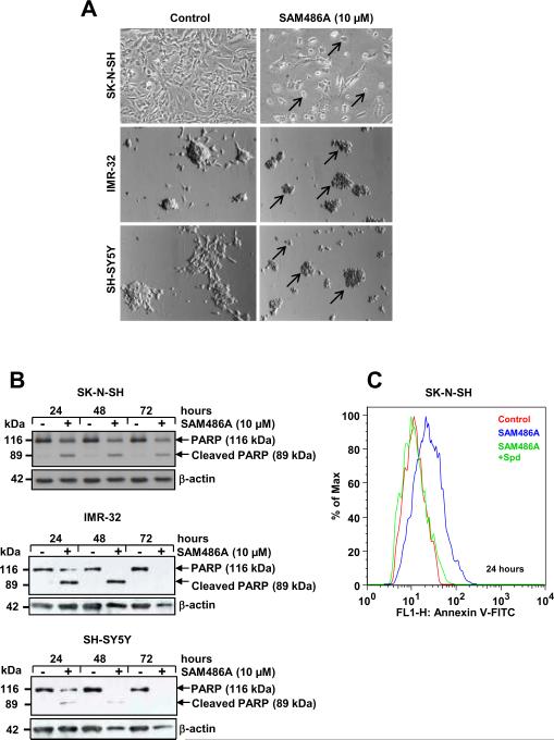
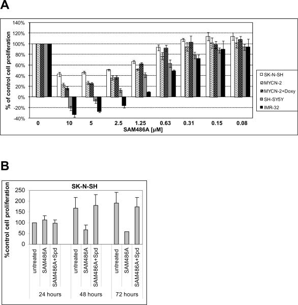
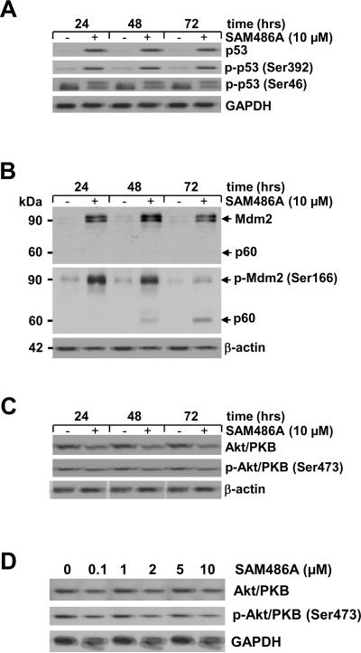
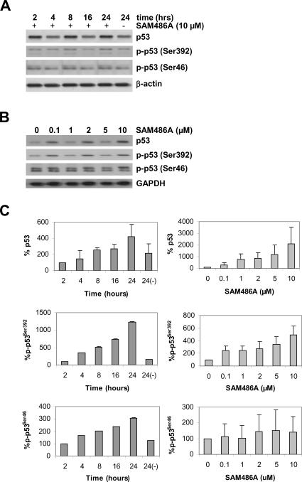
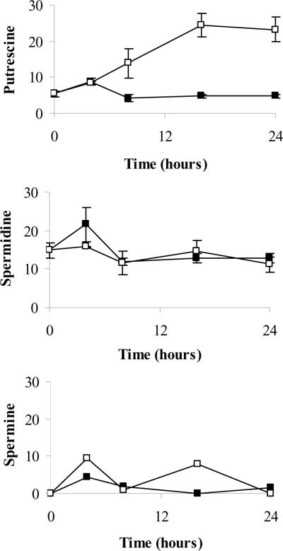
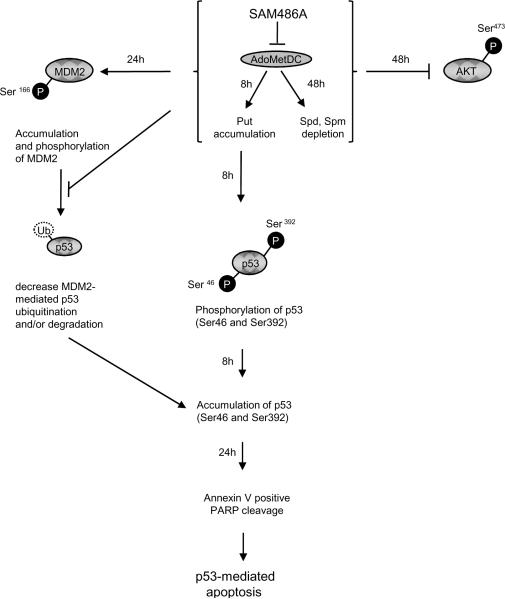
S-adenosylmethionine decarboxylase (AdoMetDC) is an essential enzyme of polyamine (PA) biosynthesis, and both AdoMetDC and PA levels are often up-regulated in cancer cells. The second-generation inhibitor SAM486A inhibits AdoMetDC enzyme activity and has been evaluated in phase II clinical cancer trials. However, little is known about the mechanism of action and potential use of this therapeutic drug in the treatment of the pediatric cancer neuroblastoma (NB). Here, we show that p53 wild-type NB cells are highly sensitive to SAM486A treatment. Most notably, SAM486A treatment resulted in the rapid accumulation of proapoptotic proteins p53 and Mdm2. Concomitant with the increase of proteins at endogenous levels, the in vivo phosphorylation of p53 at residues Ser(46)/Ser(392) and Mdm2 at residue Ser(166) was observed. Moreover, the antiapoptotic protein Akt/protein kinase B was down-regulated and also dephosphorylated at residue Ser(473) in a dose- and time-dependent manner and NB cells entered apoptotic cell death. The results presented in this study highlight the importance of PA homeostasis and provide a direct link between PA metabolism and apoptotic cell signaling pathways in p53 wild-type NB cells. PA inhibitors such as SAM486A may be effective alternative agents for the treatment of NBs with or without MYCN amplification.


Figures

Figure 1
Figure 1
SAM486A induces apoptosis in p53-wild type NB Cells. SK-N-SH, IMR-32, and SH-SY5Y cells were exposed to 10 μM SAM486A or left untreated for 24, 48, and/or 72 hours. A, representative light micrographs demonstrate the effects of SAM486A on the cell morphology (black arrows) after 72 hours. B, whole cell lysates were analyzed by Western blot for PARP cleavage, a marker for late apoptosis, after 24, 48, and 72 hours. C, SK-N-SH cells were analyzed with flow cytometry and annexin V staining, an early apoptosis marker, to confirm the induction of apoptotic cell death after 24 hours. Spd (10μM) reversed SAM486A-induced apoptosis. Similar results (without Spd control) were obtained after 48 and 72 hours (not shown). Data are representative of three independent experiments (n=3). Spd control in C (n=2).
Figure 2
Figure 2
SAM486A inhibits cell proliferation in p53-wild type NB cells. Cell proliferation rate was determined by tetrazolium-based MTS assay. A, the dose-dependent effect of SAM486A was determined in SK-N-SH, SH-SY5Y, IMR-32, and MYCN-2 cells (± doxycycline; Doxy), at 72 hours. B, cells were left untreated or exposed to 10 μM SAM486A, in the presence or absence of 10 μM Spd and 1 mM aminoguanidine, for 24, 48, and 72 hours. Data represent the mean (± SE) of three independent experiments (n=3).
Figure 3
Figure 3
Analysis of apoptosis-regulating proteins in SAM486A-treated NB cells. Whole cell lysates were obtained from SK-N-SH cells exposed to 10 μM SAM486A for 24, 48, and 72 hours or exposed to increasing concentrations of SAM486A (0, 0.1, 1, 2, 5, and 10 μM) for 72 hours, and analyzed by Western blot. Resolved proteins were transferred to PVDF membranes and sequentially probed for (A) total p53, phospho-p53 (Ser392), and phospho-p53 (Ser46), (B) total Mdm2 and phospho-Mdm2 (Ser 166), (C-D) total Akt/PKB and phospho-Akt/PKB (Ser473). SAM486A treatment increased total p53, phospho-p53 (Ser392), phopho-p53 (Ser46), total Mdm2, phospho-Mdm2 (Ser166) and decreased total Akt/PKB and phospho-Akt/PKB (Ser473). Total Akt/PKB and phospho-Akt/PKB (Ser473) also decreased in a dose-dependent manner. GAPDH or β-actin served as loading markers. Data are representative of three independent experiments (n=3).
Figure 4
Figure 4
Rapid response of p53 in SAM486A-treated NB cells. Whole cell lysates were obtained from SK-N-SH cells exposed to (A) 10 μM SAM486A for 2, 4, 8, 16, and 24 hours or (B) exposed to increasing concentrations of SAM486A (0, 0.1, 1, 2, 5, and 10 μM) for 72 hours, and analyzed by Western blot. Resolved proteins were transferred to PVDF membranes and sequentially probed for total p53, phospho-p53 (Ser392), and phospho-p53 (Ser46). SAM486A treatment increased total p53, phospho-p53 (Ser392) and phospho-p53 (Ser46) in a time-and dose-dependent manner. These effects were observed as early as 8 hours after treatment with 10 μM SAM486A, and at concentrations as low as 1-2 μM SAM486A. GAPDH or β-actin served as loading markers. C, the bands were quantified using a Bio-Rad Multi Imager and Quantity One Quantitation Software (Bio-Rad), and normalized to the untreated control group. Western blot data are representative of three independent experiments (n=3).
Figure 5
Figure 5
Intracellular PA pool determination in SAM486A-treated NB cells. SK-N-SH cells were exposed to 10 μM SAM486A or left untreated. Intracellular Put, Spd, and Spm pools were measured at 0, 4, 8, 16, and 24 hours. At 8 hours, SAM486A-treated cells had a significantly larger Put content compared to untreated cells. Put levels continued to rise in later time points. SAM486A did not have any significant effects on Spd and Spm throughout the 24 hour time course. Filled squares (■) and open squares (□) represent data collected from untreated and SAM486A-treated NB cells, respectively. Data are expressed as nmol/mg protein and represent the mean (± SE) of three independent experiments (n=3).
Figure 6
Figure 6
Schematic diagram showing proposed interplay between PAs and apoptosis-regulating proteins in p53-wild type NB cells. AdoMetDC is a key enzyme necessary for the production of dcAdoMet, a precursor in PA biosynthesis. Inhibition of AdoMetDC by SAM486A leads to the accumulation of Put as early as 8 hours after treatment. Remarkably, within the same time period, a rapid increase of native p53 protein levels was observed, suggesting that Put may be linked to p53-mediated apoptosis in these NB cells. The observed apoptosis-inducing properties of the clinically-tested SAM486A may be most effective in the treatment of NB patients with p53-wild type status, independent of their MYCN amplification status.

References

    1. Davidson NE, Hahm HA, McCloskey DE, Woster PM, Casero RA., Jr Clinical aspects of cell death in breast cancer: the polyamine pathway as a new target for treatment. Endocr Relat Cancer. 1999;6:69–73. - PubMed
    1. Levin VA, Hess KR, Choucair A, et al. Phase III randomized study of postradiotherapy chemotherapy with combination alpha-difluoromethylornithine-PCV versus PCV for anaplastic gliomas. Clin Cancer Res. 2003;9:981–90. - PubMed
    1. Levin VA, Uhm JH, Jaeckle KA, et al. Phase III randomized study of postradiotherapy chemotherapy with alpha-difluoromethylornithine-procarbazine, N-(2-chloroethyl)-N'-cyclohexyl-N-nitrosurea, vincristine (DFMO-PCV) versus PCV for glioblastoma multiforme. Clin Cancer Res. 2000;6:3878–84. - PubMed
    1. McCann PP, Pegg AE. Ornithine decarboxylase as an enzyme target for therapy. Pharmacol Ther. 1992;54:195–215. - PubMed
    1. Meyskens FL, Jr, Gerner EW. Development of difluoromethylornithine (DFMO) as a chemoprevention agent. Clin Cancer Res. 1999;5:945–51. - PubMed

Publication types

MeSH terms