

Review
doi: 10.3233/JAD-2009-1152.
Effects of stress and stress hormones on amyloid-beta protein and plaque deposition
Affiliations
- PMID: 19584430
- PMCID: PMC2905685
- DOI: 10.3233/JAD-2009-1152
Item in Clipboard
Review
Effects of stress and stress hormones on amyloid-beta protein and plaque deposition
J Alzheimers Dis.
2009.
Abstract
Growing evidence indicates that physical and psychosocial stressors, in part acting through the hypothalamic-pituitary-adrenal (HPA) axis, may accelerate the process of Alzheimer's disease (AD). In this review, we summarize recent research related to the effects of stress and stress hormones on the various disease process elements associated with AD. Specifically, we focus on the relationships among chronic stressors, HPA axis activity, amyloid-beta protein, and amyloid-beta plaque deposition in mouse models of AD. The potential mechanisms by which stress and stress-related components, especially corticotrophin-releasing factor and its receptors, influence the pathogenesis of AD are discussed.
Figures
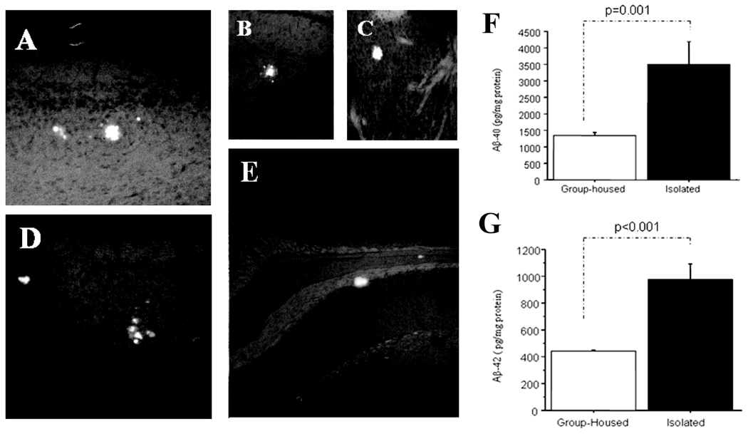
Thioflavin S fluorescence stains indicate that relatively smaller sizes of Aβ-plaques were distributed in the cortex (panel A and D), hippocampus (panel B and E), even in the stratum (panel C) [40, 80] after 6 months of isolation stress in the Tg+ mice brain, which were rare in Tg+ mice in age-matched group-housed controls. ELISA analysis indicates Aβ1–40 and Aβ1–42 levels in brain tissue were significantly increased in Aβ1–40 (panel F) and Aβ1–42 (panel G) in the 6 months isolation-stressed Tg+ mice as compared with group-housed Tg+ mice at the same age [87].
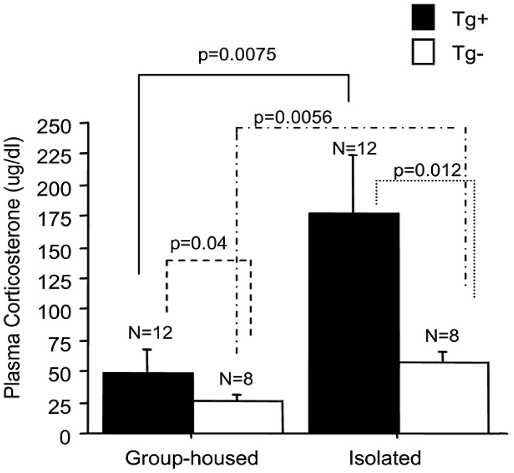
Plasma corticosterone levels in Tg+ and Tg− mice. Regardless of housing condition, plasma corticosterone levels were significantly higher in Tg+ mice than Tg− mice. Plasma corticosterone levels were dramatically increased in both Tg+ and Tg− mice after 24 weeks of isolation stress [80].
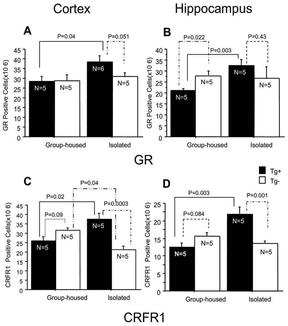
Quantitative analysis of GR and CRFR1 expression in Tg+ and Tg− mice. GR-immunoreactive cell density was significantly increased in isolated Tg+ mice as compared with group-housed Tg+ mice in the cortex and the hippocampus (A, B). CRFR1-immunoreactive cell density was significantly increased in the cortex of isolated Tg+ mice as compared with group-housed Tg+ mice and isolated Tg− mice. CRFR1-immunoreactive cell density was significantly decreased in the cortex of isolated Tg− mice as compared with group-housed Tg− mice (C). CRFR1 immunoreactive cell density was significantly increased in the hippocampus of isolated Tg+ mice as compared with group-housed Tg+ mice and isolated Tg− mice, but there was no significant change between group-housed Tg− mice and isolated Tg− mice (D) [87].

CRF was administrated by reverse microdialysis in the hippocampus of 3- to 4-month-old Tg2576 mice. (A) 100nM CRF in the microdialysis fluid resulted in an increase ISF Aβ levels at 3h after drug infusion, whereas 200nM CRF increased ISF Aβ levels immediately after drug infusion (n = 5 per group). (B) Both 100 and 200nM CRF increase ISF Aβ levels in a dose-dependent manner, reaching 138.3 = 7.027% and 171.9 = 17.83% of baseline by 12 h, respectively (P < 0.0001 and P < 0.0001, respectively). (C) Three-hour restraint stress increased ISF Aβ levels to 132 ± 6.896% compared with baseline 13h after the beginning of stress initiation (P < 0.003; n = 10 for stress). Treatment with α-helical CRF9–41 (αCRF9–41), a CRF receptor antagonist, given 30 min before restraint stress until the end of the experiment, blocked the stress-induced increase in ISF Aβ levels (P < 0.006; n = 5 for stress + αCRF9–41) [9].
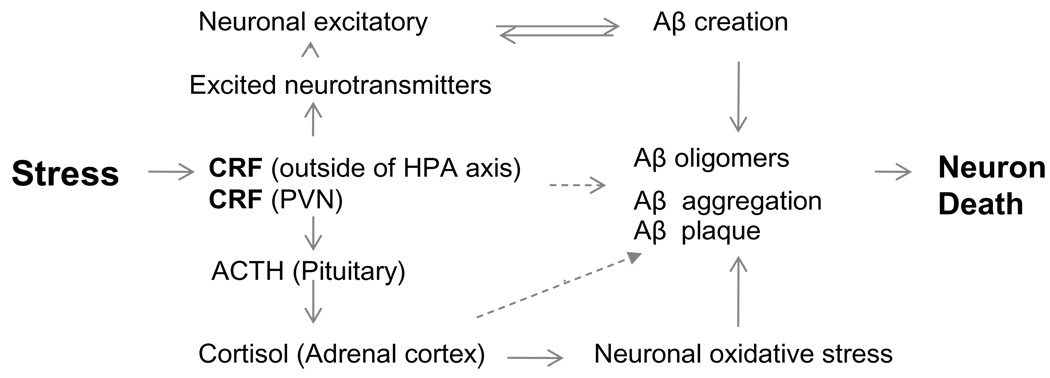
Schematic diagram depicting the theorized involvement of corticotrophin-releasing factor (CRF) in the regulation of Aβ. Stress stimulates classic hormonal and neuroregulator-like functions that may directly or indirectly influence Aβ creation and deposition.
References
-
- Arnold SE, Hyman BT, Flory J, Damasio AR, Van Hoesen GW. The topological and neuroanatomical distribution of neurofibrillary tangles and neuritic plaques in the cerebral cortex of patients with Alzheimer’s disease. Cerebral Cortex. 1991;1:103–116. - PubMed
-
- Katzman R. Alzheimer’s disease. N Engl J Med. 1986;314:964–973. - PubMed
-
- Storandt M, Grant EA, Miller JP, Morris JC. Rates of progression in mild cognitive impairment and early Alzheimer’s disease. Neurology. 2002;59:1034–1041. - PubMed
-
- Stozicka Z, Zilka N, Novak M. Risk and protective factors for sporadic Alzheimer’s disease. Acta Virol. 2007;51:205–222. - PubMed
-
- Bruandet A, Richard F, Bombois S, Maurage CA, Deramecourt V, Lebert F, Amouyel P, Pasquier F. Alzheimer’s disease with cerebrovascular disease and vascular dementia: clinical features and course compared with Alzheimer’s disease. J Neurol Neurosurg Psychiatry. 2009;80:133–139. - PubMed
Publication types
MeSH terms
Substances
Grants and funding
LinkOut - more resources
Full Text Sources
Medical
