

The gallium complex KP46 exerts strong activity against primary explanted melanoma cells and induces apoptosis in melanoma cell lines
- PMID: 19584767
- PMCID: PMC3371751
- DOI: 10.1097/CMR.0b013e32832b272d
The gallium complex KP46 exerts strong activity against primary explanted melanoma cells and induces apoptosis in melanoma cell lines
Abstract
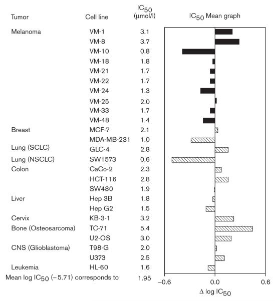
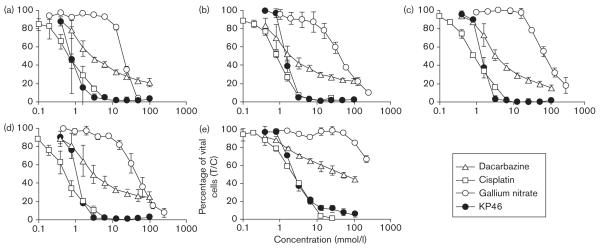
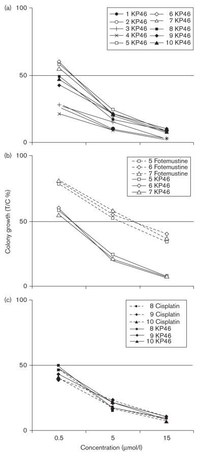
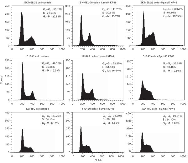
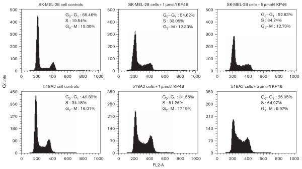
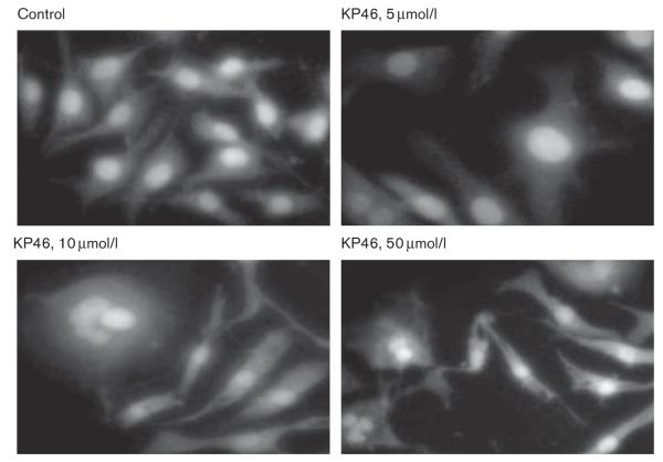
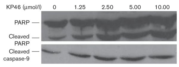
The antineoplastic properties of gallium are well documented. Owing to their robust accumulation of gallium, melanoma cells should be amenable to gallium-based anticancer drugs. With the aim of improving the disappointingly low activity of inorganic gallium salts, we have developed the orally bioavailable gallium complex KP46 [tris(8-quinolinolato)gallium(III)] that had already been successfully studied in a phase I clinical trial. To assess its therapeutic potential in malignant melanoma, its antiproliferative effects were investigated in series of human cell lines and primary explanted melanoma samples by means of the MTT [3-(4,5-dimethylthiazol-2-yl)-2,5-diphenyltetrazolium bromide] assay and the Human Tumor Cloning Assay, respectively. When compared with other cell lines, the majority of melanoma cells rank among the KP46-sensitive cell lines (50% inhibitory concentration values: 0.8-3.7 micromol/l). Clinically achievable concentrations of KP46 proved to be highly effective in melanoma cells from primary explants of cutaneous and lymph node metastases. Colony growth was inhibited in 10 of 10 specimens by 5 micromol/l KP46 (corresponding to the steady-state plasma concentration measured earlier in a study patient) and in four of 10 specimens by 0.5 micromol/l KP46. In-vitro potency of KP46 is higher than that of dacarbazine or fotemustine and comparable with that of cisplatin. The effects induced by KP46 in melanoma cell lines involve cell-cycle perturbations (S-phase arrest) and apoptosis (activation of caspase-9, PARP [poly(ADP-ribose) polymerase] cleavage, formation of apoptotic bodies). No effects on DNA secondary structure could be observed in an electrophoretic mobility shift assay using double-stranded plasmid DNA. Thus, further studies on the therapeutic applicability of KP46 in malignant melanoma are warranted.
Figures

Similar articles
-
Distinct activity of the bone-targeted gallium compound KP46 against osteosarcoma cells - synergism with autophagy inhibition.J Exp Clin Cancer Res. 2017 Apr 12;36(1):52. doi: 10.1186/s13046-017-0527-z. J Exp Clin Cancer Res. 2017. PMID: 28403890 Free PMC article.
-
Calpain-mediated integrin deregulation as a novel mode of action for the anticancer gallium compound KP46.Mol Cancer Ther. 2014 Oct;13(10):2436-49. doi: 10.1158/1535-7163.MCT-14-0087. Epub 2014 Jul 31. Mol Cancer Ther. 2014. PMID: 25082959
-
Advances in developing tris(8-quinolinolato)gallium(iii) as an anticancer drug: critical appraisal and prospects.Metallomics. 2009;1(3):193-8. doi: 10.1039/b902861g. Epub 2009 Apr 9. Metallomics. 2009. PMID: 21305117 Review.
-
The gallium complex KP46 sensitizes resistant leukemia cells and overcomes Bcl-2-induced multidrug resistance in lymphoma cells via upregulation of Harakiri and downregulation of XIAP in vitro.Biomed Pharmacother. 2022 Dec;156:113974. doi: 10.1016/j.biopha.2022.113974. Epub 2022 Nov 5. Biomed Pharmacother. 2022. PMID: 36411649
-
Gallium Complexes as Anticancer Drugs.Met Ions Life Sci. 2018 Feb 5;18:/books/9783110470734/9783110470734-016/9783110470734-016.xml. doi: 10.1515/9783110470734-016. Met Ions Life Sci. 2018. PMID: 29394029 Review.
Cited by
-
Gallium compound GaQ(3) -induced Ca(2+) signalling triggers p53-dependent and -independent apoptosis in cancer cells.Br J Pharmacol. 2012 May;166(2):617-36. doi: 10.1111/j.1476-5381.2011.01780.x. Br J Pharmacol. 2012. PMID: 22074401 Free PMC article.
-
Medical applications and toxicities of gallium compounds.Int J Environ Res Public Health. 2010 May;7(5):2337-61. doi: 10.3390/ijerph7052337. Epub 2010 May 10. Int J Environ Res Public Health. 2010. PMID: 20623028 Free PMC article. Review.
-
In Vivo Trafficking of the Anticancer Drug Tris(8-Quinolinolato) Gallium (III) (KP46) by Gallium-68/67 PET/SPECT Imaging.Molecules. 2023 Oct 22;28(20):7217. doi: 10.3390/molecules28207217. Molecules. 2023. PMID: 37894695 Free PMC article.
-
Automated synthesis of [68Ga]oxine, improved preparation of 68Ga-labeled erythrocytes for blood-pool imaging, and preclinical evaluation in rodents.Medchemcomm. 2018 Feb 1;9(3):454-459. doi: 10.1039/c7md00607a. eCollection 2018 Mar 1. Medchemcomm. 2018. PMID: 30108935 Free PMC article.
-
Anticancer Tungstenocenes with a Diverse Set of (O,O-), (O,S-) and (O,N-) Chelates-A Detailed Biological Study Using an Improved Evaluation via 3D Spheroid Models.Pharmaceutics. 2023 Jul 3;15(7):1875. doi: 10.3390/pharmaceutics15071875. Pharmaceutics. 2023. PMID: 37514061 Free PMC article.
References
-
- Van der Wall H, McLaughlin AF, Southee AE. Gallium scintigraphy in tumor diagnosis and management. In: Murray IPC, Ell PJ, editors. Nuclear medicine in clinical diagnosis and treatment. 2nd ed Vol. 2. Churchill Livingston; Edinburgh: 1998. pp. 813–829.
-
- Kirkwood JM, Myers JE, Vlock DR, Neumann R, Ariyan S, Gottschalk A, et al. Tomographic gallium-67 citrate scanning: useful new surveillance for metastatic melanoma. Ann Intern Med. 1982;97:694–699. - PubMed
-
- Kagan R, Witt T, Bines S, Mesleh G, Economou S. Gallium-67 scanning for malignant melanoma. Cancer. 1988;61:272–274. - PubMed
-
- Straus DJ. Gallium nitrate in the treatment of lymphoma. Semin Oncol. 2003;30(Suppl 5):25–33. - PubMed
-
- Einhorn L. Galium nitrate in the treatment of bladder cancer. Semin Oncol. 2003;30(Suppl 5):34–41. - PubMed
Publication types
MeSH terms
Substances
Grants and funding
LinkOut - more resources
Full Text Sources
Other Literature Sources
Medical
