. 2009 Jul;15(7):750-6.
doi: 10.1038/nm.1983. Epub 2009 Jul 5.

A small molecule blocking oncogenic protein EWS-FLI1 interaction with RNA helicase A inhibits growth of Ewing's sarcoma

Affiliations

A small molecule blocking oncogenic protein EWS-FLI1 interaction with RNA helicase A inhibits growth of Ewing's sarcoma

Hayriye V Erkizan et al. Nat Med. 2009 Jul.

Abstract

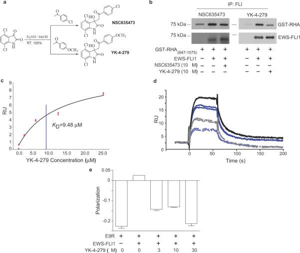
Many sarcomas and leukemias carry nonrandom chromosomal translocations encoding tumor-specific mutant fusion transcription factors that are essential to their molecular pathogenesis. Ewing's sarcoma family tumors (ESFTs) contain a characteristic t(11;22) translocation leading to expression of the oncogenic fusion protein EWS-FLI1. EWS-FLI1 is a disordered protein that precludes standard structure-based small-molecule inhibitor design. EWS-FLI1 binding to RNA helicase A (RHA) is important for its oncogenic function. We therefore used surface plasmon resonance screening to identify compounds that bind EWS-FLI1 and might block its interaction with RHA. YK-4-279, a derivative of the lead compound from the screen, blocks RHA binding to EWS-FLI1, induces apoptosis in ESFT cells and reduces the growth of ESFT orthotopic xenografts. These findings provide proof of principle that inhibiting the interaction of mutant cancer-specific transcription factors with the normal cellular binding partners required for their oncogenic activity provides a promising strategy for the development of uniquely effective, tumor-specific anticancer agents.


Figures

Figure 1
Figure 1. RHA is necessary for optimal transformation by EWS-FLI1
a. A schematic representation of RHA including the region that binds to EWS-FLI1. The E9R peptide corresponds to amino acids 823 to 832, located in the proximal of HA2 region of RHA. b. An shRNA expression vector was transfected into TC71 (ESFT) cells to reduce RHA levels. c. TC71 viability was reduced, as measured by WST reduction, following RHA shRNA expression. d. Alanine mutagenesis within E9R sequence was followed by in vitro immunoprecipitation with EWS-FLI1. The density of the GST-RHA band was measured and this graph is the average of three experiments. RHA P824A and D827A mutants have significantly lower binding to EWS-FLI1 (*p= 0.0129 and **p = 0.0034 respectively). e. Murine fibroblasts were placed in soft-agar for anchorage-independent growth assays (empty vector (W), EWS-FLI1 alone (WEF1)). f. The graph enumerates the colonies counted in three separate experiments; the difference between wild-type and mutant RHA was significant (*p= 0.0028). g. Protein expression for the fibroblasts is shown, detected with anti-FLAG (top) or anti-FLI1 (bottom). h. Densitometry of the EWS-FLI1 blot was performed using MultiGauge software.
Figure 2
Figure 2. E9R peptide prevents EWS-FLI1 binding to RHA with specific detrimental effects upon ESFT growth and transformation
a. Immunoprecipitation of GST-RHA(647-1075) using recombinant full-length EWS-FLI1 bound to a FLI1 antibody. b. Growth reduction upon E9R-P (Antennapedia-E9R) treatment (10 μM) was observed in TC32 cells but not SKNAS cells. c. E9R-P peptide uptake was tracked with FITC label (upper panels). Merged images of DAPI nuclear counter-stain (middle panels) and FITC-Ant-E9R (lower panels) showed peptide was distributed throughout the cytoplasm and nucleus of both TC32 and SKNAS cells. Scale bar equals 20 μm. d. Neither Antennapedia alone (Antp), nor a mutant of an aspartic acid residue important for RHA binding to EWS-FLI1 (E9R-D5A-P) reduced growth of TC32 cells while E9R-P reduced cell growth. e. TC71 and SKNAS cells expressed EGFP empty vector (pG), EGFP-E9R (pGE9R), EGFP with nuclear export sequence (pGC) or EGFP-E9R with nuclear export sequence (pGCE9R). Only expression of EGFP-E9R in TC71 reduced anchorage-independent growth. f. Colony numbers of three experiments are averaged with a significant reduction in only TC71 cells when expressing E9R throughout the cell (*p = 0.0012). Scale bar equals 20 μm.
Figure 3
Figure 3. Small molecule binds to EWS-FLI1 and displaces E9R from EWS-FLI1
a. NSC635437, 3-hydroxy-3-(2-oxo-2-phenyl-ethyl)-1,3-dihydro-indol-2-one was synthesized with 100% yield. Aromatic optimization produced YK-4-279 a para-methoxy derivative of NSC635437. b. EWS-FLI1 was incubated with NSC635437 (left panel) or YK-4-279 (right panel) followed by the addition of GST-RHA(647-1075). A FLI1 antibody complexed EWS-FLI1 and precipitated it from the solution. c. YK-4-279 steady state kinetics for binding to recombinant EWS-FLI1 that was immobilized on a CM5 Biacore chip. d. SPR displacement assay of 64 μM E9R alone (Black solid line) and with addition of YK-4-279 (Grey dashed line); 32 μM E9R alone (Dark blue solid line) and with addition of YK-4-279 (Light blue dashed line). e. Fluorescent polarization indicated the binding of 3.2 μM of FITC-E9R to EWS-FLI1, which was competitively inhibited by increasing concentrations of YK-4-279.
Figure 4
Figure 4. YK-4-279 reduces EWS-FLI1 functional activity
a. TC32 cells were treated with YK-4-279 and resolved protein lysates were immunoblotted for co-precipitated RHA (top), EWS-FLI1 (middle), or total RHA (bottom). b. Luciferase reporter assay of EWS-FLI1 responsive NR0B1 promoter showed YK-4-279 dose-dependent (18-hour treatment) reduction in the promoter activity in COS7 cells. c. Protein lysates from transfected cells showed expression of EWS-FLI1. d. YK-4-279 treated TC32 cell lysates (treated for 14 hours) were blotted for cyclin D1 and actin.
Figure 5
Figure 5. YK-4-279 is potent and specific inhibitor of ESFT
a. TC32 cells were treated with a dose range of YK-4-279 and NSC635437. Cell number measured by MTT or WST reduction after seven days in culture. b. TC32 and HEK-293 (non-transformed, lacking EWS-FLI1) were treated similarly to (a). c. Primary ESFT explant cell lines GUES1 and ES925 were treated for 3 days with YK-4-279. d. Cell lines expressing EWS-FLI1 were compared to non-EWS-FLI1 malignant cell lines following 3 days in culture to establish the IC50 using WST assay. e. Caspase 3 activity of a panel of ESFT (TC32, TC71, A4573, and ES925), malignant non-EWS-FLI1 expressing (MCF-7, MDA-MB-231, PC3, ASPC1, COLO-PL), and non-transformed cells (HEK-293, HFK, and HEC) were measured. Graph plots level of fluorescence in treated divided by untreated lysate. f. Arrows indicated apoptotic nuclear fragmentation after 50 μM YK-4-279 treated ESFT (TC32) and non-transformed cells (HEK-293, HFK, and HEC). Scale bar equals 200 μm.
Figure 6
Figure 6. YK-4-279 inhibited the growth of ESFT xenograft tumors
Xenografts were established with injection of either ESFT (CHP-100 or TC71) or Prostate cancer (PC3) cells. a. CHP-100 intramuscular xenografts (arrow indicates when tumors were palpable) received DMSO (n=4; open circles) or 1.5 mg YK-4-279 (n=5; triangles) (p=0.016, by t-Test comparison). Single experiment growth curves depicted are representative of five independent experiments. b. PC3 subcutaneous xenografts (arrow indicates when tumors were palpable) were treated as CHP-100 cells (a) (n=5 per group, representative of 3 independent experiments). c. Overall response of ESFT xenografts (TC71, open symbols, and CHP-100, closed symbols) to YK-4-279 (1.5 mg/dose). Tumor volumes at day 14 after treatment initiation compared across 5 experiments (DMSO n = 19, YK-4-279 n = 25, p < 0.0001, by Mann-Whitney test). d. Tumors from the mice in (a) were analyzed for activation of caspase-3 activity using immunohistochemistry. e. Caspase-3 positive cells were counted (n>500 in 3 high-power-fields) in 4 separately stained slides for each group (p = 0.041).

References

    1. Mitelman F, Johansson B, Mertens F. The impact of translocations and gene fusions on cancer causation. Nat Rev Cancer. 2007;7:233–245. - PubMed
    1. French CA, et al. Midline carcinoma of children and young adults with NUT rearrangement. J Clin Oncol. 2004;22:4135–4139. - PubMed
    1. Helman LJ, Meltzer P. Mechanisms of sarcoma development. Nat Rev Cancer. 2003;3:685–694. - PubMed
    1. Poppe B, et al. Expression analyses identify MLL as a prominent target of 11q23 amplification and support an etiologic role for MLL gain of function in myeloid malignancies. Blood. 2004;103:229–235. - PubMed
    1. Carroll M, et al. CGP 57148, a tyrosine kinase inhibitor, inhibits the growth of cells expressing BCR-ABL, TEL-ABL, and TEL-PDGFR fusion proteins. Blood. 1997;90:4947–4952. - PubMed

Publication types

MeSH terms