. 2010 Jan;18(1):73-80.
doi: 10.1038/ejhg.2009.117.

Specific epigenetic alterations of IGF2-H19 locus in spermatozoa from infertile men

Affiliations

Specific epigenetic alterations of IGF2-H19 locus in spermatozoa from infertile men

Céline Chalas Boissonnas et al. Eur J Hum Genet. 2010 Jan.

Abstract

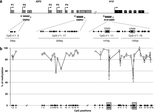
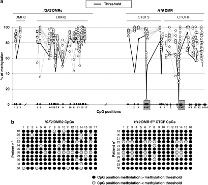
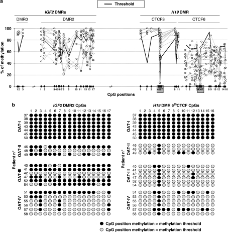
DNA methylation marks, a key modification of imprinting, are erased in primordial germ cells and sex specifically re-established during gametogenesis. Abnormal epigenetic programming has been proposed as a possible mechanism compromising male fertility. We analysed by pyrosequencing the DNA methylation status of 47 CpGs located in differentially methylated regions (DMRs), the DMR0 and DMR2 of the IGF2 gene and in the 3rd and 6th CTCF-binding sites of the H19 DMR in human sperm from men with normal semen and patients with teratozoospermia (T) and/or oligo-astheno-teratozoospermia (OAT). All normal semen samples presented the expected high global methylation level for all CpGs analysed. In the teratozoospermia group, 11 of 19 patients presented a loss of methylation at variable CpG positions either in the IGF2 DMR2 or in both the IGF2 DMR2 and the 6th CTCF of the H19 DMR. In the OAT group, 16 of 22 patients presented a severe loss of methylation of the 6th CTCF, closely correlated with sperm concentration. The methylation state of DMR0 and of the 3rd CTCF was never affected by the pathological status of sperm samples. This study demonstrates that epigenetic perturbations of the 6th CTCF site of the H19 DMR might be a relevant biomarker for quantitative defects of spermatogenesis in humans. Moreover, we defined a methylation threshold sustaining the classification of patients in two groups, unmethylated and methylated. Using this new classification of patients, the observed intrinsic imprinting defects of spermatozoa appear not to impair significantly the outcome of assisted reproductive technologies.


Figures

Figure 1
Figure 1
(a) Structural characteristics of the human IGF2–H19 locus. The IGF2 and H19 genes contain 9 exons and 5 exons, respectively (grey and black numbered squares). The transcription start sites and promoters used are indicated by arrows. Regions of differential methylation (DMRs) are shown as black (methylated allele) and white (unmethylated allele) boxes (P=paternal, M=maternal). For the IGF2 gene, two regions were analysed: IGF2 DMR0 (3 CpGs) and IGF2 DMR2 (17 CpGs). For the H19 DMR, two of the seven CTCF binding sites and their surrounding CpGs are analysed: the 3rd CTCF (11CpGs) and the 6th CTCF (16 CpGs) binding sites. CpGs corresponding to the two CTCF-binding sites are highlighted by grey squares. For each DMR, the number of CpGs analysed, their relative positions (filled circles) and the length of the amplified sequence in base pairs (bp) are indicated. (b) Methylation pattern at the IGF2-H19 locus in normozoospermic patients (NZ, n=17). The methylation at each CpG position is expressed in percentage as mean±standard deviation. The CpGs 4–7 of the 3rd and of the 6th CTCF-binding site of the H19 DMR are highlighted.
Figure 2
Figure 2
(a) Methylation pattern at the IGF2-H19 locus in teratozoospermic patients. From the methylation results of the NZ group, a mean level of methylation was calculated for each CpG position. A methylation threshold (black line) is defined as the mean value minus two SD. The methylation threshold delimits a subgroup of teratozoospermic patients (n=11) exhibiting a decrease of the DNA methylation level at specific CpG positions in the IGF2 DMR2 and the 6th CTCF-binding site of the H19 DMR. The CpG positions are represented by open circles and each line represents an individual sample analysis. Only the dots underneath the black line indicate a significant reduction of DNA methylation. (b) Alternative representation for Figure 2a. Using the methylation threshold, the closed circles represent methylated CpGs while the grey circles represents CpGs with a methylation level significantly different from the NZ group (P<0.05). CpG positions are indicated vertically (CpG 1–17 for the IGF2 DMR2 and CpGs 1–16 for the 6th CTCF-binding site of the H19 DMR). The patients are numbered and represented in lines. Eleven teratozoospermia patients showed an altered methylation status of CpG positions in the IGF2 DMR2 and 6 of them showed also alterations in the 6th CTCF-binding site of the H19 DMR.
Figure 3
Figure 3
Methylation pattern of IGF2-H19 locus in oligo-asthenoteratozoospermic patients. (a) The methylation threshold (black line) delimits a subgroup of OAT patients (n=15) exhibiting a drastic decrease of methylation level of H19 DMR (6th CTCF-binding site) associated with a loss of methylation at CpG positions in the IGF2 DMR2. The CpG positions are represented by open circles and each line represents an individual sample analysis. Only the dots underneath the black line indicate a significant reduction of DNA methylation. (b) Alternative representation for Figure 3a using the OAT subgroups as defined by sperm concentration. The closed circles represent methylated CpGs, whereas the grey circles represent CpGs with a methylation level significantly different from the NZ group (P<0.05). CpG positions are indicated vertically (CpG 1-17 for IGF2 DMR2 and CpGs 1-16 for H19 DMR). The patients are numbered and represented in lines.
Figure 4
Figure 4
Relationship between the DNA methylation level of the 6th CTCF-binding site in the H19 DMR and sperm concentration. (NZ: normozoospermia group; T: teratozoospermia group; OAT: oligo-asthe no-teratozoospermia group).

References

    1. Reik W, Dean W, Walter J. Epigenetic reprogramming in mammalian development. Science. 2001;293:1089–1092. - PubMed
    1. Brandeis M, Ariel M, Cedar H. Dynamics of DNA methylation during development. Bioessays. 1993;11:709–713. - PubMed
    1. Constancia M, Pickard B, Kelsey G, Reik W. Imprinting mechanisms. Genome Res. 1998;8:881–900. - PubMed
    1. La Salle S, Mertineit C, Taketo T, et al. Windows for sex-specific methylation marked by DNA methyltransferase expression profiles in mouse germ cells. Dev Biol. 2004;268:403–415. - PubMed
    1. Kerjean A, Dupont JM, Vasseur C, et al. Establishment of the paternal methylation imprint of the human H19 and MEST/PEG1 genes during spermatogenesis. Hum Mol Genet. 2000;9:2183–2187. - PubMed

Publication types

Substances