

MicroRNAs involved in tumor suppressor and oncogene pathways: implications for hepatobiliary neoplasia
- PMID: 19585622
- PMCID: PMC2721015
- DOI: 10.1002/hep.23010
MicroRNAs involved in tumor suppressor and oncogene pathways: implications for hepatobiliary neoplasia
Abstract
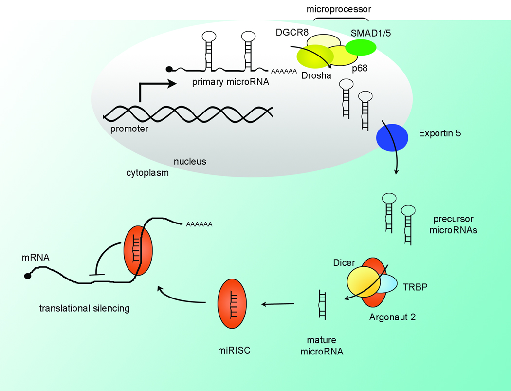
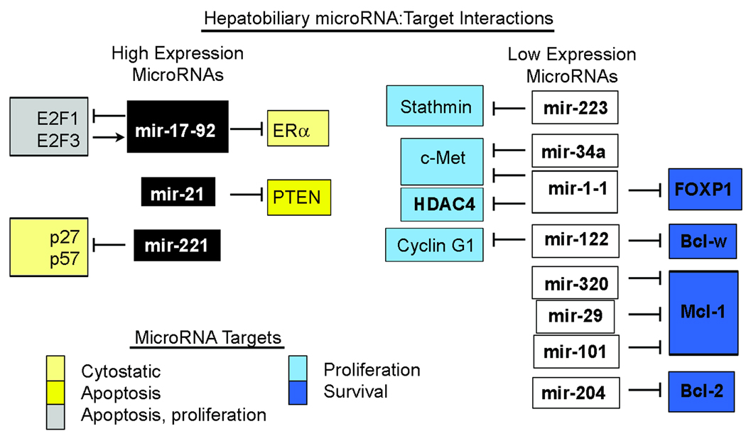
MicroRNAs are a class of small regulatory RNAs that function to modulate protein expression. This control allows for fine-tuning of the cellular phenotype, including regulation of proliferation, cell signaling, and apoptosis; not surprisingly, microRNAs contribute to liver cancer biology. Recent investigations in human liver cancers and tumor-derived cell lines have demonstrated decreased or increased expression of particular microRNAs in hepatobiliary cancer cells. Based on predicted and validated protein targets as well as functional consequences of altered expression, microRNAs with decreased expression in liver tumor cells may normally aid in limiting neoplastic transformation. Conversely, selected microRNAs that are up-regulated in liver tumor cells can promote malignant features, contributing to carcinogenesis. In addition, microRNAs themselves are subject to regulated expression, including regulation by tumor suppressor and oncogene pathways. This review will focus on the expression and function of cancer-related microRNAs, including their intimate involvement in tumor suppressor and oncogene signaling networks relevant to hepatobiliary neoplasia.
Figures
Similar articles
-
Impact of microRNAs on regulatory networks and pathways in human colorectal carcinogenesis and development of metastasis.BMC Genomics. 2013 Aug 29;14:589. doi: 10.1186/1471-2164-14-589. BMC Genomics. 2013. PMID: 23987127 Free PMC article.
-
AGR2: The Covert Driver and New Dawn of Hepatobiliary and Pancreatic Cancer Treatment.Biomolecules. 2024 Jun 23;14(7):743. doi: 10.3390/biom14070743. Biomolecules. 2024. PMID: 39062458 Free PMC article. Review.
-
MicroRNAs: small but potent oncogenes or tumor suppressors.Curr Opin Investig Drugs. 2006 Jun;7(6):560-4. Curr Opin Investig Drugs. 2006. PMID: 16784027 Review.
-
The malignant phenotype-associated microRNA in gastroenteric, hepatobiliary and pancreatic carcinomas.Expert Opin Biol Ther. 2010 Dec;10(12):1693-701. doi: 10.1517/14712598.2010.532482. Epub 2010 Oct 26. Expert Opin Biol Ther. 2010. PMID: 20977295 Review.
-
MicroRNA-548a-5p promotes proliferation and inhibits apoptosis in hepatocellular carcinoma cells by targeting Tg737.World J Gastroenterol. 2016 Jun 21;22(23):5364-73. doi: 10.3748/wjg.v22.i23.5364. World J Gastroenterol. 2016. PMID: 27340352 Free PMC article.
Cited by
-
Sevoflurane Inhibits Proliferation, Invasion, but Enhances Apoptosis of Lung Cancer Cells by Wnt/β-catenin Signaling via Regulating lncRNA PCAT6/miR-326 Axis.Open Life Sci. 2020 Apr 10;15:159-172. doi: 10.1515/biol-2020-0017. eCollection 2020. Open Life Sci. 2020. PMID: 33987473 Free PMC article.
-
Exploration of genome-wide circulating microRNA in hepatocellular carcinoma: MiR-483-5p as a potential biomarker.Cancer Epidemiol Biomarkers Prev. 2013 Dec;22(12):2364-73. doi: 10.1158/1055-9965.EPI-13-0237. Epub 2013 Oct 14. Cancer Epidemiol Biomarkers Prev. 2013. PMID: 24127413 Free PMC article.
-
MiR-148a attenuates paclitaxel resistance of hormone-refractory, drug-resistant prostate cancer PC3 cells by regulating MSK1 expression.J Biol Chem. 2010 Jun 18;285(25):19076-84. doi: 10.1074/jbc.M109.079525. Epub 2010 Apr 20. J Biol Chem. 2010. PMID: 20406806 Free PMC article.
-
MicroRNA-2053 overexpression inhibits the development and progression of hepatocellular carcinoma.Oncol Lett. 2019 Aug;18(2):2043-2049. doi: 10.3892/ol.2019.10501. Epub 2019 Jun 20. Oncol Lett. 2019. PMID: 31423276 Free PMC article.
-
From 'omics' to complex disease: a systems biology approach to gene-environment interactions in cancer.Cancer Cell Int. 2010 Apr 26;10:11. doi: 10.1186/1475-2867-10-11. Cancer Cell Int. 2010. PMID: 20420667 Free PMC article.
References
Publication types
MeSH terms
Substances
Grants and funding
LinkOut - more resources
Full Text Sources
Medical