. 2009 Jul;10(7):522-7.
doi: 10.1631/jzus.B0820295.

Features of intervertebral disc degeneration in rat's aging process

Affiliations

Features of intervertebral disc degeneration in rat's aging process

Yin-Gang Zhang et al. J Zhejiang Univ Sci B. 2009 Jul.

Abstract

Objective: The age-related change is important part of degenerative disc disease. However, no appropriate animal model or objective evaluation index is available. This study aimed to investigate the features of intervertebral disc degeneration in aging process of rats.

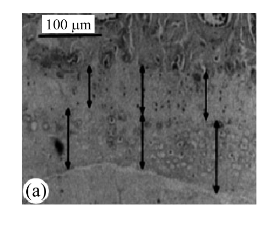
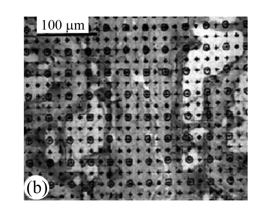
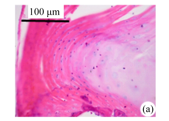
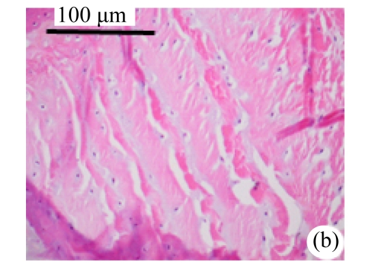
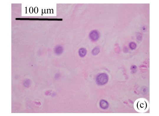
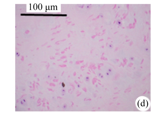
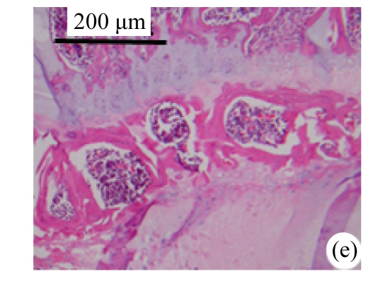
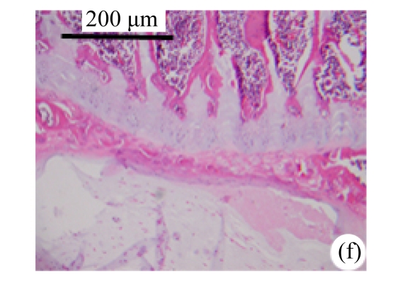
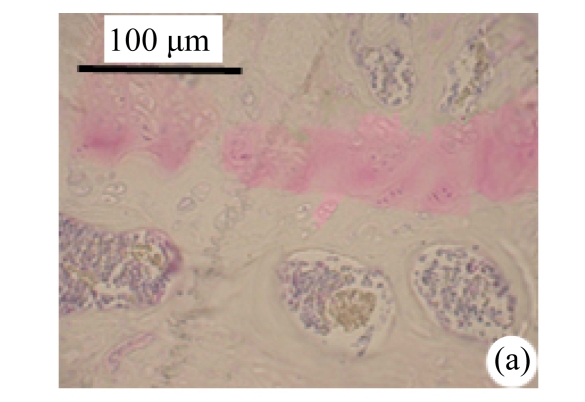
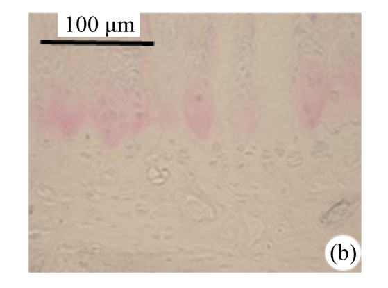
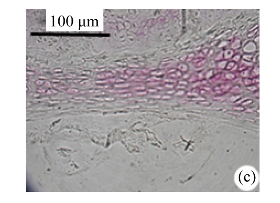
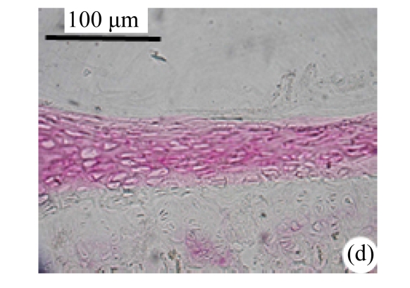
Methods: 22-month-old Sprague-Dawley (SD) rats were used as spontaneously occurring intervertebral disc degeneration models and 6-month-old rats as young controls. Expression of collagen types II and X was measured by immunohistochemistry. Degenerations of intervertebral discs were scored according to Miyamoto's method. Numbers and areas of afferent vascular buds were measured. The thicknesses of non-calcified and calcified layers were measured and statistically analyzed.

Results: There were less collagen type II expression and more collagen type X expression in the calcified layer of the cartilage endplates and nucleus pulposus in the rats of the aged group than in the young control. There were fewer and smaller afferent vascular buds in the rats of the aged group than in the young control group. The ratio of the non-calcified to the calcified layers in the rats of the aged group significantly decreased, compared with that of the young control group (P<0.01).

Conclusion: Rats can spontaneously establish intervertebral disc age-related degeneration. The expression of collagen types II and X, numbers and areas of afferent vascular buds, the ratio of the non-calcified to the calcified layers, and water and glycosaminoglycan contents in the nucleus pulposus are sensitive indexes of intervertebral disc degeneration.


Figures

Fig.1
Fig.1
Block diagram of calculation of afferent vascular bud relative area and cartilage endplate (H&E staining) (a) Vascular area was analyzed by square method and the percentage of vascular area to the whole was calculated; (b) Tidemark was used as separatrix, the thicknesses of calcified layer and non-calcified layer in front 1/3, middle point, and post 1/3 of median sagittal plane in nucleus pulposus were measured, and average values were calculated
Fig.1
Fig.1
Block diagram of calculation of afferent vascular bud relative area and cartilage endplate (H&E staining) (a) Vascular area was analyzed by square method and the percentage of vascular area to the whole was calculated; (b) Tidemark was used as separatrix, the thicknesses of calcified layer and non-calcified layer in front 1/3, middle point, and post 1/3 of median sagittal plane in nucleus pulposus were measured, and average values were calculated
Fig.2
Fig.2
Change of intervertebral disc histomorphology between young controls and aged rats The annular fibrosus arranged regularly and more cells were seen in young controls (a) than in aged rats (b). Dead cells in the nucleus gelatinosus were not observed in young controls (c), but were obvious in aged rats (d). Obviously more vascular buds were seen in young controls than in aged rats. Non-clarify layers of young controls (e) were significantly thicker than those of the aged group (f)
Fig.2
Fig.2
Change of intervertebral disc histomorphology between young controls and aged rats The annular fibrosus arranged regularly and more cells were seen in young controls (a) than in aged rats (b). Dead cells in the nucleus gelatinosus were not observed in young controls (c), but were obvious in aged rats (d). Obviously more vascular buds were seen in young controls than in aged rats. Non-clarify layers of young controls (e) were significantly thicker than those of the aged group (f)
Fig.3
Fig.3
Expression of intervertebral disc collagen types II and X between the young and aged groups Expression of collagen type II in the cartilage endplate-calcified layer increased in young rats (a), but decreased or lost in the aged group (b). Expression of collagen type X in the non-calcified layer of the cartilage endplate in young rats (c) was obviously smaller than that of the aged group (d)
Fig.3
Fig.3
Expression of intervertebral disc collagen types II and X between the young and aged groups Expression of collagen type II in the cartilage endplate-calcified layer increased in young rats (a), but decreased or lost in the aged group (b). Expression of collagen type X in the non-calcified layer of the cartilage endplate in young rats (c) was obviously smaller than that of the aged group (d)

References

    1. Ahsan R, Tajima N, Chosa E, Sugamata M, Sumida M, Hamada M. Biochemical and morphological changes in herniated human intervertebral disc. J Orthop Sci. 2001;6(6):510–518. doi: 10.1007/s007760100006. - DOI - PubMed
    1. Ala-Kokko L. Genetic risk factors for lumbar disc disease. Ann Med. 2002;34(1):42–47. doi: 10.1080/078538902317338634. - DOI - PubMed
    1. Annunen S, Paassilta P, Lohiniva J, Perälä M, Pihlajamaa T, Karppinen J, Tervonen O, Kröger H, Lähde S, Vanharanta H, et al. An allele of COL9A2 associated with intervertebral disc disease. Science. 1999;285(5426):409–412. doi: 10.1126/science.285.5426.409. - DOI - PubMed
    1. Antoniou J, Steffen T, Nelson F, Winterbottom N, Hollander AP, Poole RA, Aebi M, Alini M. The human lumbar intervertebral disc: evidence for changes in the biosynthesis and denaturation of the extracellular matrix with growth, maturation, ageing, and degeneration. Journal of Clinical Investigation. 1996;98(4):996–1003. doi: 10.1172/JCI118884. - DOI - PMC - PubMed
    1. Boos N, Nerlich AG, Wiest I, von der Mark K, Aebi M. Immunolocalization of type X collagen in human lumbar intervertebral discs during ageing and degeneration. Histochem Cell Biol. 1997;108(6):471–480. doi: 10.1007/s004180050187. - DOI - PubMed

Publication types