Review
. 2009 Jul;135(4):638-77.
doi: 10.1037/a0015849.

Source monitoring 15 years later: what have we learned from fMRI about the neural mechanisms of source memory?

Affiliations
Review

Source monitoring 15 years later: what have we learned from fMRI about the neural mechanisms of source memory?

Karen J Mitchell et al. Psychol Bull. 2009 Jul.

Abstract

Focusing primarily on functional magnetic resonance imaging (fMRI), this article reviews evidence regarding the roles of subregions of the medial temporal lobes, prefrontal cortex, posterior representational areas, and parietal cortex in source memory. In addition to evidence from standard episodic memory tasks assessing accuracy for neutral information, the article considers studies assessing the qualitative characteristics of memories, the encoding and remembering of emotional information, and false memories, as well as evidence from populations that show disrupted source memory (older adults, individuals with depression, posttraumatic stress disorder, or schizophrenia). Although there is still substantial work to be done, fMRI is advancing understanding of source memory and highlighting unresolved issues. A continued 2-way interaction between cognitive theory, as illustrated by the source monitoring framework (M. K. Johnson, S. Hashtroudi, & D. S. Lindsay, 1993), and evidence from cognitive neuroimaging studies should clarify conceptualization of cognitive processes (e.g., feature binding, retrieval, monitoring), prior knowledge (e.g., semantics, schemas), and specific features (e.g., perceptual and emotional information) and of how they combine to create true and false memories.


Figures

Figure 1
Figure 1
Anatomical relationships between various temporal regions. (A) Three-D figure of the human brain (frontal lobes are to the left), with amygdala and hippocampus indicated. (B) View of temporal regions from below; top of the figure is the front of the brain. (C) Cut-away of medial temporal lobe structures as viewed from the front (adapted with permission from Figure 1A of Dolcos, F., LaBar, K. S., & Cabeza, R. [2004]. Interaction between the amygdala and the medial temporal lobe memory system predicts better memory for emotional events. Neuron, 42, 855–863). A-amygdala, E-entorhinal cortex, FG-fusiform gyrus, H-hippocampus, ITG-inferior temporal gyrus, MTG-middle temporal gyrus, PR-perirhinal cortex (sometimes referred to in the literature as anterior parahippocampal gyrus), PH-parahippocampal cortex (sometimes referred to in the literature as posterior parahippocampal gyrus), TP-temporal pole.
Figure 2
Figure 2
Schematic of the relationship between MTL regions with summary of several current hypotheses regarding their functions. MTL-medial temporal lobes; PR-perirhinal cortex; PH-parahippocampal cortex. Superscripts refer to: 1Davachi (2006); 2Mayes et al. (2007); 3Diana et al. (2007); 4Eichenbaum et al. (2007); 5Awipi and Davachi (2008).
Figure 3
Figure 3
Lateral (left) and medial (right) views of the cortex. Numbers indicate approximate Brodmann Areas (BA). Abbreviations for regions (areas are approximate) are: AG-angular gyrus; CC-corpus collosum; CG-cingulate gyrus; Cu-cuneus; FG-fusiform gyrus; FP-frontal pole; IFG-inferior frontal gyrus; IOG-inferior occipital gyrus; IPS- intraparietal sulcus; ITG-inferior temporal gyrus; LG-lingual gyrus; LOG-lateral occipital gyrus; MdFG-medial frontal gyrus; MFG-middle frontal gyrus; MTG-middle temporal gyrus; OrbG-orbital gyrus; PCC-posterior cingulate cortex; PCu-precuneus; PHG-parahippocampal gyrus; PrCG-precentral gyrus; PoCG-postcentral gyrus; RSC-retrosplenial cortex; SFG-superior frontal gyrus; SOG-superior occipital gyrus; SMG-supramarginal gyrus; SPL-superior parietal lobule; STG-superior temporal gyrus; TP-temporal pole. In addition, entorhinal cortex (not labeled) is primarily comprised of BA 28, 34; perirhinal cortex-BA 35, 36.
Figure 4
Figure 4
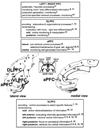
Summary of some hypotheses about functional specificity of prefrontal cortex (PFC) in source memory, and example references, as described in text. Nomenclature for anatomical regions varies historically and between labs, but generally, PFC areas involved in source memory include: Medial (hatch lines in axial view) and lateral (speckled in axial view) areas of anterior PFC (aPFC; primarily BA 10); aPFC is also sometimes referred to as fronto-polar cortex, or rostral PFC. Dorsolateral PFC (DLPFC = primarily dorsal BA 10, BAs 46, 9, but some investigators also include BA 8 and dorsal BA 6; mid-DLPFC = primarily BAs 9, 46; posterior DLPFC = primarily BA 9). Ventrolateral PFC (VLPFC = primarily BAs 47, 45, 44, but some investigators also include BAs 11 and ventral 6; anterior VLPFC = primarily BA 47; mid-VLPFC = primarily BA 45; posterior VLPFC = primarily BA 44); VLPFC is also sometimes referred to as inferior frontal cortex (IFC). The inferior frontal sulcus is usually taken as the boundary between DLPFC and VLPFC (e.g., Kuhl & Wagner, 2009). Superscripts refer to: 1Blumenfeld and Ranganath (2007); 2Buckner and Wheeler (2001); 3Cabeza et al. (2003); 4Dobbins and Han (2006); 5Dobbins and Wagner (2005); 6Kelley et al. (1998); 7Lepage et al. (2000); 8McDermott et al. (1999); 9Mitchell et al., 2004; 10Mitchell et al. (2008); 11Nolde, Johnson, and Raye, (1998); 12Petrides (2002); 13Ranganath and Blumenfeld, 2008; 14Raye et al. (2000); 15Rugg et al. (2002; see also, Hayama et al., 2008); 16Simons et al. (2008); 17Simons, Gilbert, et al., (2005); 18Vinogradov et al. (2006; see also Vinogradov et al., 2008); 19Wagner, Poldrack, et al. (1998).

Comment on

  • Source monitoring.
    Johnson MK, Hashtroudi S, Lindsay DS. Johnson MK, et al. Psychol Bull. 1993 Jul;114(1):3-28. doi: 10.1037/0033-2909.114.1.3. Psychol Bull. 1993. PMID: 8346328 Review.

References

    1. Achim AM, Weiss AP. No evidence for a differential deficit of reality monitoring in schizophrenia: A meta-analysis of the associative memory literature. Cognitive Neuropsychiatry. 2008;13:369–384. - PubMed
    1. Ackil JK, Zaragoza MS. Memorial consequences of forced confabulation: Age differences in susceptibility to false memories. Developmental Psychology. 1998;34:1358–1372. - PubMed
    1. Addis DR, Moscovitch M, Crawley AP, McAndrews MP. Recollective qualities modulate hippocampal activation during autobiographical memory retrieval. Hippocampus. 2004;14:752–762. - PubMed
    1. Aggleton JP, Brown MW. Episodic memory, amnesia, and the hippocampal-anterior thalamic axis. Behavioral and Brain Sciences. 1999;22:425–489. - PubMed
    1. Aleman A, Hijman R, De Haan EHF, Kahn RS. Memory impairment in schizophrenia: a meta-analysis. American Journal of Psychiatry. 1999;156:1358–1366. - PubMed

MeSH terms