. 2009 Sep 4;284(36):24213-22.
doi: 10.1074/jbc.M109.000745. Epub 2009 Jul 8.

Checkpoint kinase ATR promotes nucleotide excision repair of UV-induced DNA damage via physical interaction with xeroderma pigmentosum group A

Affiliations

Checkpoint kinase ATR promotes nucleotide excision repair of UV-induced DNA damage via physical interaction with xeroderma pigmentosum group A

Steven M Shell et al. J Biol Chem. .

Abstract

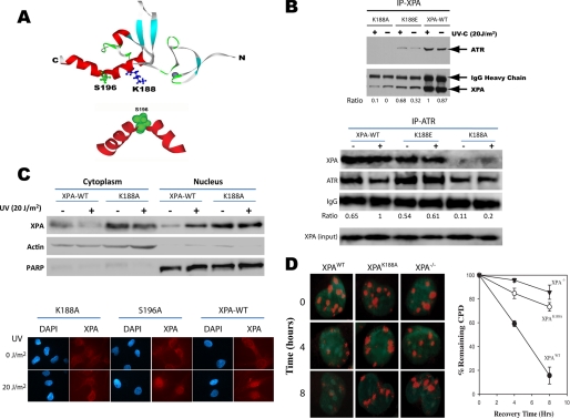
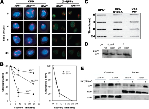
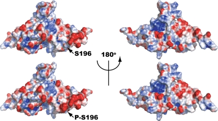
In response to DNA damage, eukaryotic cells activate a series of DNA damage-dependent pathways that serve to arrest cell cycle progression and remove DNA damage. Coordination of cell cycle arrest and damage repair is critical for maintenance of genomic stability. However, this process is still poorly understood. Nucleotide excision repair (NER) and the ATR-dependent cell cycle checkpoint are the major pathways responsible for repair of UV-induced DNA damage. Here we show that ATR physically interacts with the NER factor Xeroderma pigmentosum group A (XPA). Using a mass spectrometry-based protein footprinting method, we found that ATR interacts with a helix-turn-helix motif in the minimal DNA-binding domain of XPA where an ATR phosphorylation site (serine 196) is located. XPA-deficient cells complemented with XPA containing a point mutation of S196A displayed a reduced repair efficiency of cyclobutane pyrimidine dimers as compared with cells complemented with wild-type XPA, although no effect was observed for repair of (6-4) photoproducts. This suggests that the ATR-dependent phosphorylation of XPA may promote NER repair of persistent DNA damage. In addition, a K188A point mutation of XPA that disrupts the ATR-XPA interaction inhibits the nuclear import of XPA after UV irradiation and, thus, significantly reduced DNA repair efficiency. By contrast, the S196A mutation has no effect on XPA nuclear translocation. Taken together, our results suggest that the ATR-XPA interaction mediated by the helix-turn-helix motif of XPA plays an important role in DNA-damage responses to promote cell survival and genomic stability after UV irradiation.


Figures

FIGURE 1.
FIGURE 1.
XPA-ATR complex preparation. A, U2OS-ATR cells were treated with increasing amounts of doxycycline for 24 h to induce expression of the FLAG-ATR construct. Then, cell lysates were mixed with anti-FLAG IgG, and immunoprecipitated proteins were analyzed by Western blot using anti-ATR monoclonal IgG. Western blot analysis indicates that as the concentration of doxycycline increases, the amount of FLAG-ATR expression also increases, whereas immunoprecipitation of the FLAG-tagged protein results in a single full-length protein. B, FLAG-ATR protein was immunoprecipitated and washed with increasing concentrations of NaCl solution to remove bound proteins. ATRIP forms a tight complex with ATR and is efficiently removed by washing with 1 m NaCl. The ATRIP/ATR ratios were normalized to the ratio at zero salt concentration. C, recombinant His6-XPA was immobilized on nickel-nitrilotriacetic acid beads and modified with increasing amounts of NHS-biotin (0, 0.1, 0.25, 0.5, and 1 mm) before or after the addition of FLAG-ATR lysates. XPA modified with 1 mm NHS-biotin before the addition of FLAG-ATR lysate prevents formation of the XPA-ATR complex; however, modification after complex formation does not affect the protein-protein interaction. A nonspecific (NS) protein band was also observed during the immunoprecipitation even though no XPA was added to the beads, indicating that the protein interacts with the nickel matrix and not with XPA or ATR (data not shown). Because of the nonspecific nature of the band, it was used as a loading control for these experiments. D, FLAG-ATR purified by immunoprecipitation was mixed with His6-XPA (lane 3 and 4) and modified with NHS-biotin (lane 4). As control, anti-FLAG antibody was mixed with the recombinant XPA in the absence of ATR protein (lane 5). Protein bands were excised and digested with trypsin for mass spectrometry analysis.
FIGURE 2.
FIGURE 2.
MALDI-TOF analysis of biotin-modified XPA. A, a typical MALDI-TOF mass spectrum of peptide fragments resulting from trypsin digestion of biotin modified XPA. B, summary of MALDI-TOF results in the context of the XPA primary structure. The sequence of N-terminal His6 tag added for purification of the recombinant protein is shown. The His6 tag residue numbering begins with −1 and continues backward to the N terminus. The initial methionine residue of the XPA sequence is labeled +1. Amino acid sequences corresponding to tryptic peptide fragments detected by MALDI-TOF are depicted in bold. The lysine residues affected by NHS-biotin treatment are indicated by arrows.
FIGURE 3.
FIGURE 3.
MALDI-TOF analysis of lysine protection in the XPA-ATR complex. A, top and middle spectra show free XPA and the XPA-ATR complex treated with NHS-biotin. The bottom spectrum shows untreated free XPA. The peak corresponding to XPA tryptic peptide fragment amino acids 184–189 containing a single modified Lys at position 188 is indicated. This peak is detected in free XPA samples and is significantly diminished in the XPA-ATR complex. B, unlike Lys-188, lysine residue 31 is modified in both free XPA and the XPA-ATR complex. Peaks C1, C2, and C3 are unmodified peptide fragments of XPA and serve as internal controls.
FIGURE 4.
FIGURE 4.
Residue Lys-188 is required for XPA-ATR complex formation, XPA nuclear import upon UV damage, and NER. A, ribbon diagram of XPA minimum DNA-binding domain (PDB code 1D4U), indicating that the identified lysine residue Lys-188 (shown in blue) is located in the helix-turn-helix motif-containing ATR phosphorylation site serine 196. B, point mutations were generated in the pcDNA-XPA expression construct generating alanine (K188A) and glutamic acid (K188E) substitutions. The mutated constructs as well as wild-type XPA were stably expressed in XPA−/− cells, and their effects on the XPA-ATR interaction were investigated by coimmunoprecipitation (IP). The K188A mutant protein was unable to coimmunoprecipitate ATR or vice versa from lysates generated from UV-irradiated or un-irradiated cells. The K188E mutant maintained the interaction between XPA and ATR and exhibited a similar UV-induced pattern as seen for XPA-WT. The relative amounts of the co-immunoprecipitated ATR were estimated by its ratio to those of the immunoprecipitated protein that were normalized to the loading control IgG. C, XPA cells complemented with wild-type XPA and XPA-K188A were subjected to subcellular fractionation and immunofluorescence microscopy analysis. The specificity of the fractionation assay is demonstrated by the presence and absence of cytoplasm-specific and nucleus-specific proteins β-actin and PARP (poly(ADP-ribose) polymerase), respectively, in the cytosol and nucleus. D, cells were irradiated with UV of 50 J/m2 through isopore filters to induce localized DNA damage and then fixed at the indicated times for immunofluorescence analysis with anti-CPD antibody. Nuclei containing at least one well defined CPD focus were counted as a percentage of total DAPI-stained nuclei and plotted versus time post-irradiation. At least 50 DAPI-stained nuclei were randomly chosen for the quantification at each time point.
FIGURE 5.
FIGURE 5.
Effects of XPA phosphorylation on repair of cyclobutane pyrimidine dimers. Recombinant pcDNA constructs containing wild-type XPA or XPA-S196A cDNA were stably expressed in XPA−/− cells. A, cells were grown on coverslips and UV-irradiated at 20 J/m2 through isopore filters to induce localized DNA damage followed by immunofluorescence staining with anti-(6-4)PPs and anti-CPD antibodies at the indicated time points. B, nuclei containing at least one DNA damage focus were counted as a percentage of total nuclei and plotted versus time post-irradiation. At least 50 DAPI-stained nuclei were randomly counted for each time point. C, genomic DNA was isolated from cells complemented with wild-type or phosphorylation-deficient XPA after UV-C irradiation. The DNA was then immobilized on nylon membranes, and total CPDs were detected using mouse-anti-CPD. D, cells expressing recombinants XPA-WT and XPA-K188A, respectively, were UV-irradiated and then subjected to Western blot analysis of phosphorylated and intact XPA. E, subcellular distribution of XPA-WT and XPA-S196A after UV-C irradiation was determined by cellular fractionation. β-Actin and PARP (poly(ADP-ribose) polymerase), cytoplasm- and nucleus-specific proteins, respectively, demonstrate the specificity of the assay and serve as loading controls.
FIGURE 6.
FIGURE 6.
Modeling analysis of the effects on surface charge distribution from phosphorylation of XPA at serine residue 196. A surface representation of the DNA-binding domain of XPA (PDB code 1D4U) is shown at the left with the electrostatic field mapped in red (negative) and blue (positive). A computation model of the DNA-binding domain of XPA phosphorylated at serine 196 is shown at the right with the same mapping of the electrostatic field. The circle highlights the region of the protein near serine 196 where a large net increase in negative charge is associated with phosphorylation.

Similar articles

Cited by

References

    1. Sancar A., Lindsey-Boltz L. A., Unsal-Kaçmaz K., Linn S. (2004) Annu. Rev. Biochem. 73, 39–85 - PubMed
    1. Park C. J., Choi B. S. (2006) FEBS J. 273, 1600–1608 - PubMed
    1. Riedl T., Hanaoka F., Egly J. M. (2003) EMBO J. 22, 5293–5303 - PMC - PubMed
    1. Kraemer K. H., Lee M. M., Andrews A. D., Lambert W. C. (1994) Arch. Dermatol. 130, 1018–1021 - PubMed
    1. Kraemer K. H., Lee M. M., Scotto J. (1987) Arch. Dermatol. 123, 241–250 - PubMed

Publication types

MeSH terms

Substances