. 2009 Aug 1;183(3):1767-79.
doi: 10.4049/jimmunol.0802167. Epub 2009 Jul 8.

Inflamed lymphatic endothelium suppresses dendritic cell maturation and function via Mac-1/ICAM-1-dependent mechanism

Affiliations

Inflamed lymphatic endothelium suppresses dendritic cell maturation and function via Mac-1/ICAM-1-dependent mechanism

Simona Podgrabinska et al. J Immunol. .

Abstract

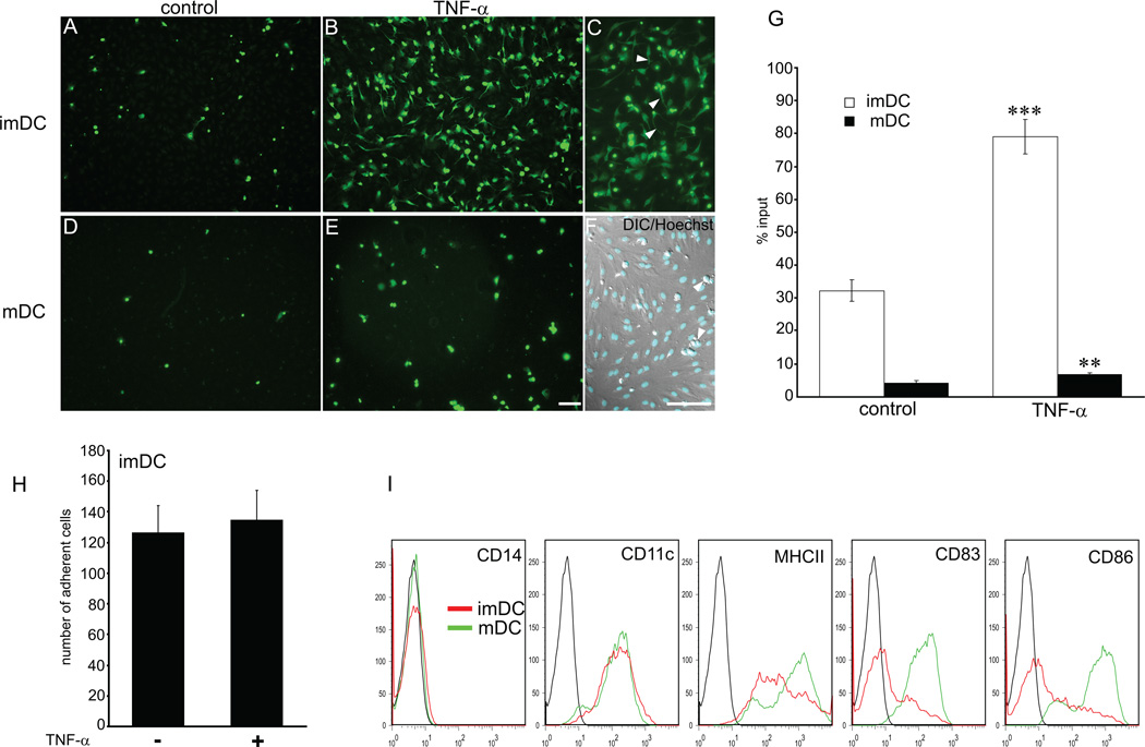
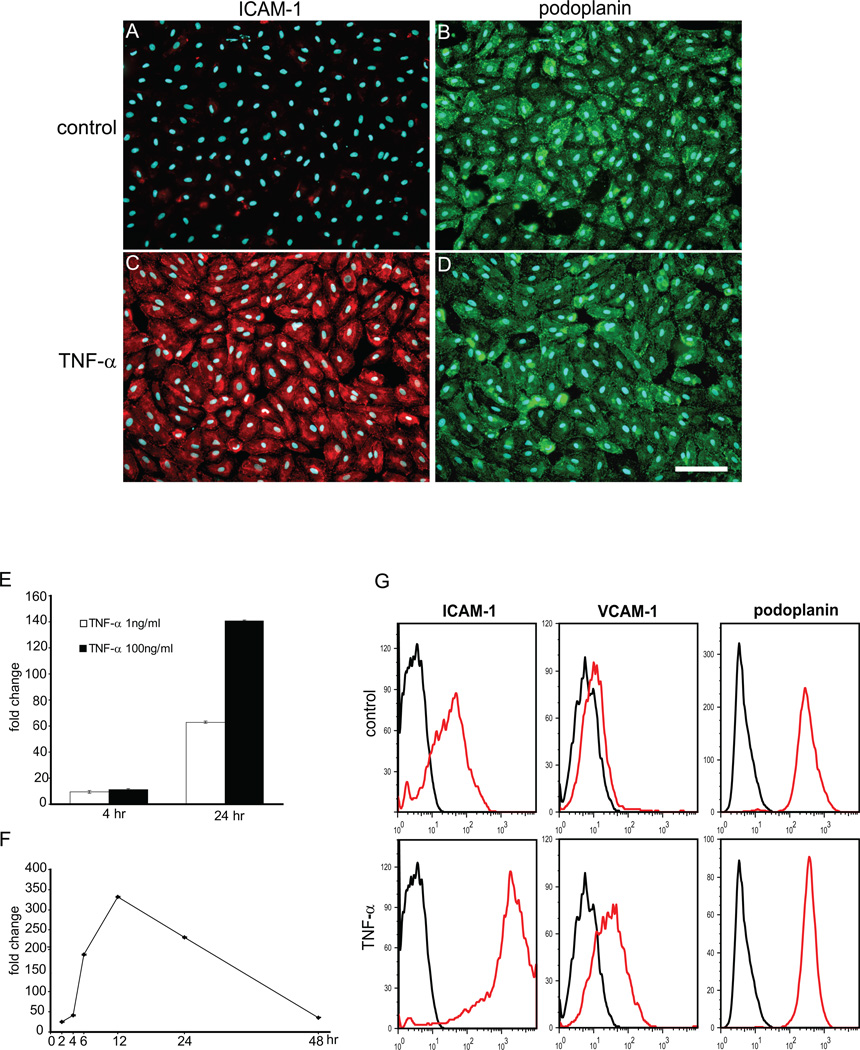
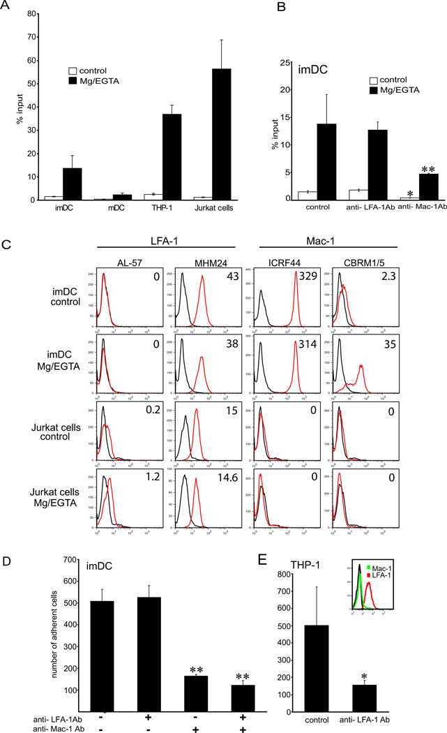
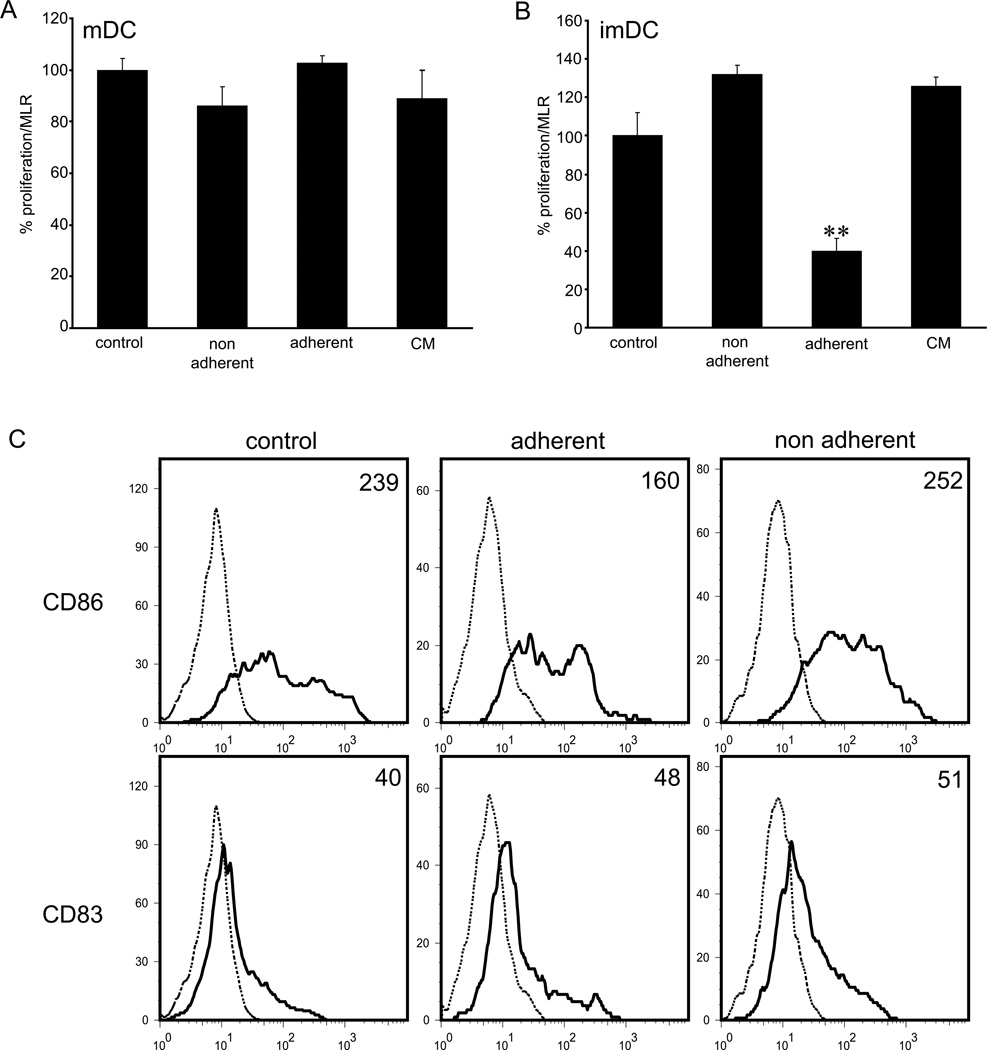
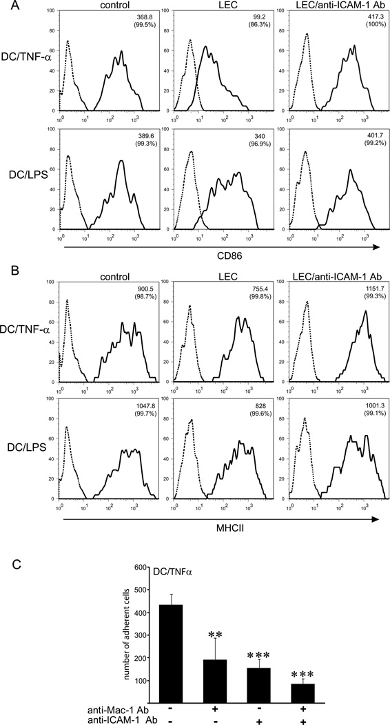
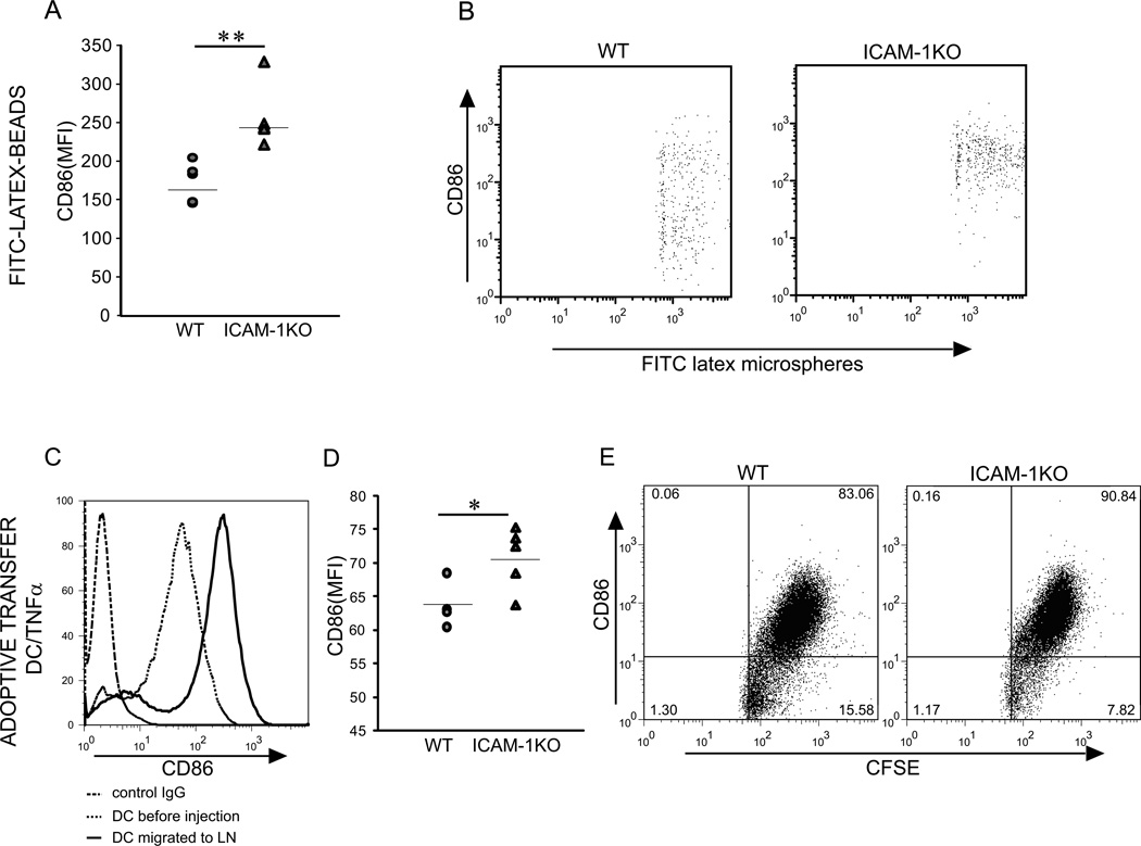
The lymphatic system is essential for the generation of immune responses by facilitating immune cell trafficking to lymph nodes. Dendritic cells (DCs), the most potent APCs, exit tissues via lymphatic vessels, but the mechanisms of interaction between DCs and the lymphatic endothelium and the potential implications of these interactions for immune responses are poorly understood. In this study, we demonstrate that lymphatic endothelial cells (LECs) modulate the maturation and function of DCs. Direct contact of human monocyte-derived DCs with an inflamed, TNF-alpha-stimulated lymphatic endothelium reduced expression of the costimulatory molecule CD86 by DCs and suppressed the ability of DCs to induce T cell proliferation. These effects were dependent on adhesive interactions between DCs and LECs that were mediated by the binding of Mac-1 on DCs to ICAM-1 on LECs. Importantly, the suppressive effects of the lymphatic endothelium on DCs were observed only in the absence of pathogen-derived signals. In vivo, DCs that migrated to the draining lymph nodes upon inflammatory stimuli, but in the absence of a pathogen, showed increased levels of CD86 expression in ICAM-1-deficient mice. Together, these data demonstrate a direct role of LECs in the modulation of immune response and suggest a function of the lymphatic endothelium in preventing undesired immune reactions in inflammatory conditions.


Figures

Figure 1
Figure 1. DC adhesion to lymphatic endothelial cells is increased with TNFα
Adhesion of imDC and LPS/mDC to control (A, D) and TNFα treated LECs (B–F) was examined in a static adhesion assay. imDCs showed higher affinity for LEC in steady-state (A) and in inflammatory condition (B) than mDC (D and E). Upon contact with TNF-α/LEC (1ng/ml, 24 hr) imDCs changed phenotype from round to dendritic (B, C). (C) Arrowheads point to protrusions. In contrast, mDCs did not change morphology upon contact with TNF-α/LECs (E). (F) Nuclear staining with Hoechst overlaid on Nomarsky image shows DCs (arrowheads) adhering to confluent LEC. (Bar = 100 µm). (G) DC adhesion was quantified by FACS and expressed as percentage of input cells. (H) Binding of imDCs to TNF-α/LECs in presence or absence of TNFα in the media. Note no difference in the adhesion levels. (I) Phenotype of DCs employed in the adhesion assays was determined by FACS just before the experiment. Data shown are representative of 4 independent experiments. Statistical significance was determined with Student’s t-test. **P<0.01, ***P<0.001.
Figure 2
Figure 2. Expression of ICAM-1 on LECs in vitro
(A–D) LECs were cultured in presence or absence of TNF-α (2 ng/ml) and expression of ICAM-1(red) and podoplanin (green) was examined by double immunofluorescent staining. Note high expression levels of ICAM-1 on TNF-α/LECs (C), but not on control cells (A). Podoplanin expression was uniform in both conditions (B, D). (E, F) qRT-PCR showing dose-response (E) and time-course (F) of ICAM-1 mRNA expression in TNF-α/LECs. For the time-course, TNF-α was added at 1ng/ml. (G) Surface expression of ICAM-1 and VCAM on control and TNF-α-treated LECs was analyzed by FACS. Data shown are representative of 2 independent experiments.
Figure 3
Figure 3. imDCs bind ICAM-1 via Mac-1
(A) Cell adhesion to ICAM-1Fc immobilized on the plate with and without Mg2+/EGTA stimulation. Note that imDCs adhere to ICAM-1 more than LPS/mDCs. (B) imDCs were preincubated with blocking antibodies to LFA-1 or Mac-1 and assayed for adhesion to ICAM-1Fc (20µg/ml). Binding of control and Mg2+/EGTA-activated imDCs is inhibited by blocking Mac-1, but not LFA-1. Data shown are representative of 3 experiments, performed in triplicates. (C) To examine the activation state of β2 integrins on imDCs, cells were incubated with antibodies recognizing active conformation of LFA-1 (AL-57 Ab) or Mac-1 (CBRM1/5 Ab) or control antibodies (MHM24 and ICRF44, respectively), and analyzed by FACS. Jurkat cells were used as a positive control for LFA-1 activation and negative control for Mac-1. Data are expressed as mean fluorescence intensity (MFI), and are representative of 2 independent experiments. (D) Adhesion of imDCs to TNFα-treated LECs preblocked with function-blocking antibodies to LFA-1 and Mac-1. Note that Mac-1 Ab inhibits adhesion of imDCs, but LFA-1 Ab does not. Adhesion of THP-1 cells, which express activated LFA-1, but not Mac-1 was inhibited when blocking LFA-1 (E). Data shown are representative of 3 experiments, performed in triplicates. Appropriate isotype controls were used in all experiments (see Methods). Statistical significance was determined with Student’s t-test. *P<0.05, **P<0.01.
Figure 4
Figure 4. Adhesion of imDCs to TNFα-stimulated LECs suppresses T-cell proliferation
(A, B) LPS/mDCs or imDCs were co-cultured with TNFα/LECs, equal numbers of non-adherent and adherent cells were collected and assayed in MLR. DCs maintained in the culture media or in the LEC-conditioned media (CM) were used as controls. Note significant reduction of T-cell proliferation only when adherent imDCs were employed in the assay. Data shown represent the 1:10 ratio of DCs to T-cells. (C) FACS analysis of imDCs for surface expression of CD83 and CD86 prior to MLR shows reduced expression of CD86. Data are expressed as mean fluorescence intensity (MFI). Data shown are representative of 3 independent experiments. Statistical significance was determined with Student’s t-test. **P<0.01.
Figure 5
Figure 5. Interaction of TNFα-matured DCs with ICAM-1 expressed by LECs decreases expression of CD86 in the absence of antigen
(A, B) Dendritic cells matured with LPS (DC/LPS) or with TNFα (DC/TNFα) were co-cultured for 12hr with TNFα/LECs or TNFα/LECs preincubated with the anti-ICAM-1 blocking antibody. DCs incubated in the same culture media for 12hr were used as a control. All DCs from co-cultures (adherent and non-adherent) were analyzed by FACS. (A) Histograms show that CD86 is downregulated on DC/TNFα upon contact with LECs, but not on DC/LPS. This effect was reversed by adding a blocking antibody to ICAM-1. (B) MHCII expression on DC/TNFα or DC/LPS in co-cultures with TNFα/LECs. Note modest change of MHCII expression, when compared to CD86. Data are expressed as a mean fluorescence intensity (MFI); percentage of positive cells is indicated in brackets. (C) Adhesion of DC/TNFα to TNFα-treated LECs preblocked with function-blocking antibodies to ICAM-1, Mac-1 or both. Data shown are representative of 3 independent experiments. Statistical significance was determined with Student’s t-test. **P<0.01, ***P<0.001.
Figure 6
Figure 6. The effect of ICAM-1 deficiency on CD86 expression by DCs migrated into regional lymph nodes
(A, B) FITC-latex microspheres were injected into the footpads of WT and ICAM-1 KO mice (n=5). After 2 days draining LNs were collected (3 per mouse), pooled and phenotype of migrated DCs with beads was analyzed by FACS. (A) Quantitative comparison of CD86 surface expression by migrated DCs. (B) Representative plots depicting entire population of microsphere-bearing cells recovered from the lymph nodes. (C–E) Adoptive transfer of bone marrow-derived TNFα-matured DCs into the back skin of WT and ICAM-1 KO mice (n=5). (C) FACS analysis of CD86 expression on DCs immediately before injection and on DCs which migrated into the LNs of WT mice. (D) Comparison of CD86 surface expression on migrated DCs in WT and ICAM-1 KO mice. (E) Representative plots depicting entire population of CFSE-labeled DCs recovered from the lymph nodes. Data shown are representative of at least 2 experiments. Statistical significance was determined with Student’s t-test. *P<0.05, **P<0.01.
Figure 7
Figure 7. Expression of ICAM-1 on collecting lymphatic vessels and on lymphatic sinuses in mouse lymph node
(A–F) Confocal analysis of ICAM-1 localization on lymphatic endothelium of a collecting vessel 20 hr after injection of Latex beads into the mouse footpad. Triple immunofluorescent staining for LYVE-1 (green), ICAM-1 (red) and α-SMA (blue) shows expression of ICAM-1 on the apical (luminal) side of the vessel (arrows) and on the basal side of the endothelium (arrowheads). (G–L) Double immunofluorescent staining for LYVE-1 (green) and ICAM-1 (red) of control lymph nodes (G–I) and lymph nodes drainig footpad 20 hr after injection of 200 ng TNFα (J–L) Cell nuclei are counterstained with Hoechst (grey in A–C; blue in H, K). L designates a leukocyte expressing ICAM-1, but not LYVE-1. Scale bars: A–F: 7.5 µm; G–L: 50 µm.

References

    1. Pepper MS, Skobe M. Lymphatic endothelium: morphological, molecular and functional properties. J.Cell Biol. 2003;163:209–213. - PMC - PubMed
    1. Angeli V, Randolph GJ. Inflammation, lymphatic function, and dendritic cell migration. Lymphat Res Biol. 2006;4:217–228. - PubMed
    1. Olszewski WL. The innate reaction of the human skin lymphatic system to foreign and self-antigens. Lymphat Res Biol. 2005;3:50–57. - PubMed
    1. Pugh CW, MacPherson GG, Steer HW. Characterization of nonlymphoid cells derived from rat peripheral lymph. J Exp Med. 1983;157:1758–1779. - PMC - PubMed
    1. Mackay CR, Marston WL, Dudler L. Naive and memory T cells show distinct pathways of lymphocyte recirculation. J Exp Med. 1990;171:801–817. - PMC - PubMed

Publication types