. 2010 Mar;20(3):661-71.
doi: 10.1093/cercor/bhp134. Epub 2009 Jul 8.

Transforming growth factor beta promotes neuronal cell fate of mouse cortical and hippocampal progenitors in vitro and in vivo: identification of Nedd9 as an essential signaling component

Affiliations

Transforming growth factor beta promotes neuronal cell fate of mouse cortical and hippocampal progenitors in vitro and in vivo: identification of Nedd9 as an essential signaling component

Tanja Vogel et al. Cereb Cortex. 2010 Mar.

Abstract

Transforming Growth Factor beta (Tgfbeta) and associated signaling effectors are expressed in the forebrain, but little is known about the role of this multifunctional cytokine during forebrain development. Using hippocampal and cortical primary cell cultures of developing mouse brains, this study identified Tgfbeta-regulated genes not only associated with cell cycle exit of progenitors but also with adoption of neuronal cell fate. Accordingly, we observed not only an antimitotic effect of Tgfbeta on progenitors but also an increased expression of neuronal markers in Tgfbeta treated cultures. This effect was dependent upon Smad4. Furthermore, in vivo loss-of-function analyses using Tgfbeta2(-/-)/Tgfbeta3(-/-) double mutant mice showed the opposite effect of increased cell proliferation and fewer neurons in the cerebral cortex and hippocampus. Gata2, Runx1, and Nedd9 were candidate genes regulated by Tgfbeta and known to be involved in developmental processes of neuronal progenitors. Using siRNA-mediated knockdown, we identified Nedd9 as an essential signaling component for the Tgfbeta-dependent increase in neuronal cell fate. Expression of this scaffolding protein, which is mainly described as a signaling molecule of the beta1-integrin pathway, was not only induced after Tgfbeta treatment but was also associated with morphological changes of the Nestin-positive progenitor pool observed upon exposure to Tgfbeta.


Figures
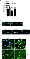
Figure 1.
Figure 1.
Primary cortical and hippocampal neurons isolated from E14.5 and E16.5 mouse embryonic brains express Tgfβs and their receptors and are responsive to TGFβ treatment. (A) MLEC/PAI–luciferase assay with conditioned medium from DIV4 cortical and hippocampal cultures. Data as mean ± SEM (n = 3). (B) RT-PCR reveals that cortical and hippocampal neurons express Tgfβ receptors I and II, as well as Tgfβ isoforms 1, 2, and 3. (C) Hippocampal neurons activate Smad proteins upon TGFβ stimulation. Smad1/2/3 immunoreactivity in control untreated cells was localized diffusely in the cytoplasm, whereas TGFβ treatment caused a translocation of Smad proteins in the nucleus. Upper panels: DIV4, lower panels: DIV12. Scale bars = 50 μm.
Figure 2.
Figure 2.
TGFβ promotes exit from the cell cycle. (A) Hippocampal and cortical cultures were treated for 6 days with TGFβ, labeled with BrdU (20 nM) in the last 24 h, and stained for BrdU and Ki67, detecting decreased numbers of proliferating progenitors (n = 3), P values: ***P < 0.001, **P < 0.01, student's t-test. (B) Determination of cells leaving the cell cycle within the first 48 h of TGFβ exposure. Treatment started at DIV2; after 24 h, BrdU was added and after another 24 h, cells were fixed and costained for BrdU and Ki67. BrdU incorporation had not changed at this time point, but there was an increase in the number of cells quitting the cell cycle (QF) and a corresponding decrease of cells that reenter the cell cycle (CF) (all n = 3), P values: ***P < 0.001, **P < 0.01, student's t-test. (C) PI-FACS analysis of apoptotic cells after short-time (DIV2–DIV4) and long-time (DIV2–DIV8) treatment with TGFβ (n = 3).
Figure 3.
Figure 3.
TGFβ promotes neuronal differentiation from hippocampal and cortical progenitors in a Smad-dependent way. (A) Immunohistochemical appearance of HuC/D and NeuN expressing neurons, Nestin-positive progenitors, and GFAP-positive glia cells in hippocampal cultures after 6 days of TGFβ treatment compared with untreated control cells. Scale bars = 50 μm. (B,C) Quantification of markers reveals an increase of neurons and a decrease in progenitors in TGFβ-treated cultures compared with untreated controls. The number of glia cells remains unchanged. Data are presented as a percentage of the total cell number. Statistical significance was determined by student's t-test with P values: ***P < 0.001, **P < 0.01. (D) Quantification of HuC/D-positive neurons after 6-day stimulation of hippocampal cells with Tgfβ2 and Tgfβ3, respectively, at 8 DIV. (E) Influence of the duration of the TGFβ pulse and the age of the cultures on the TGFβ-induced neurogenetic effect in hippocampal cultures. A short TGFβ pulse of 24 h was sufficient to enhance neuronal differentiation (2,5,7) in the same way as a long pulse of 6 days over the entire cultivation period (1,4,6). TGFβ was also able to promote a neuronal cell fate in older cultures from DIV5 (4,5) at a similar rate as in cultures from DIV2 (1,2). In cultures where TGFβ treatment was started on DIV8 (6,7), there was less increase in the number of HuC/D neurons. Data are presented as percent difference of HuC/D-positive cells of the TGFβ-treated cultures based on the untreated controls and given as mean ± SEM (n = 3). (F) Quantification of HuC/D-positive cells under control conditions and after blocking Tgfβ signaling with Alk4,5,7 inhibitor SB431542. (G,H) TGFβ-mediated neuronal differentiation was abolished after knockdown of Smad4 by lentiviral transduction of cortical (G) and hippocampal (H) cultures. HuC/D-positive neurons were counted after transduction with Smad4 shRNA or nontarget shRNA control with and without TGFβ treatment (n = 3).
Figure 4.
Figure 4.
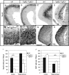
Increased proliferation and decreased neuronal differentiation in Tgfβ2/Tgfβ3 double mutant mice. (AD) Expression of NeuN in control cortex (A) and hippocampus (C), and Tgfβ2/Tgfβ3 double mutant cortex (B) and hippocampus (D) from E14.5 littermates. Scale bar = 50 μm. (E–H) Expression of Ki67 in control (E) and in Tgfβ2/Tgfβ3 double mutant cerebral cortex (F), in control (G) and in Tgfβ2/Tgfβ3 double mutant hippocampus (H). Scale bar E,F = 50 μm, G,H = 100 μm. (I, J) Quantification of NeuN (I) and Ki67 (J) positive cells in the cerebral cortex and hippocampus of age-matched control and Tgfβ2/Tgfβ3 double mutants (n = 2, P value: * P < 0.05, one-tailed student's t-test).
Figure 5.
Figure 5.
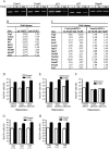
TGFβ-promoted neuronal differentiation depends on the expression of Nedd9. (A) Semiquantitative RT-PCR of cell cycle regulating genes after TGFβ stimulation of hippocampal cultures at 4DIV for 2 and 24 h. (B) Real-time RT-PCR of different Tgfβ-regulated genes using cDNA from hippocampal cultures at 4DIV. Given are fold changes after 2 and 24 h of Tgfβ treatment, respectively. (C) Real-time RT-PCR of different Tgfβ-regulated genes using cDNA from hippocampal cultures at 12DIV. Given are fold changes after 2 and 24 h of Tgfβ treatment, respectively. Cells were transduced with a nontarget shRNA control lentivirus or with a Smad4 shRNA lentivirus to assess Smad4-dependent gene regulation. (DH) shRNA-mediated knockdown of target genes showed that TGFβ-promoted neuronal differentiation was independent of Gata2 (D) and Runx1 (E). No increased neuronal differentiation was observed in cultures transduced with Nedd9 shRNA lentivirus (F). Cultures of hippocampal or cortical cells from Ctgf mutant mice (−/−) showed increased neuronal differentiation after Tgfβ treatment in similar ranges as control mice (+/+ and +/−) (G,H).
Figure 6.
Figure 6.
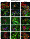
Morphological changes in Nestin-positive progenitors upon TGFβ treatment. (A–E) Preparations of hippocampal cells contain clusters of Nestin-positive progenitors that have a different morphology in untreated control cultures and also in cultures treated with Alk4,5,7-inhibitor SB431542 compared with TGFβ-treated samples. Immunohistochemical characterization with Nestin and (A) Ki67, (B) GFAP, (C) PSA-NCam, (D) Dcx, and (E) Nedd9. Scale bars = 50 μm.

References

    1. Abe M, Harpel JG, Metz CN, Nunes I, Loskutoff DJ, Rifkin DB. An assay for transforming growth factor-beta using cells transfected with a plasminogen activator inhibitor-1 promoter-luciferase construct. Anal Biochem. 1994;216:276–284. - PubMed
    1. Abramova N, Charniga C, Goderie SK, Temple S. Stage-specific changes in gene expression in acutely isolated mouse CNS progenitor cells. Dev Biol. 2005;283:269–281. - PubMed
    1. Aquino JB, Marmigere F, Lallemend F, Lundgren TK, Villar MJ, Wegner M, Ernfors P. Differential expression and dynamic changes of murine NEDD9 in progenitor cells of diverse tissues. Gene Expr Patterns. 2008;8:217–226. - PubMed
    1. Brionne TC, Tesseur I, Masliah E, Wyss-Coray T. Loss of TGF-beta 1 leads to increased neuronal cell death and microgliosis in mouse brain. Neuron. 2003;40:1133–1145. - PubMed
    1. Chapuis J, Moisan F, Mellick G, Elbaz A, Silburn P, Pasquier F, Hannequin D, Lendon C, Campion D, Amouyel P, et al. Association study of the NEDD9 gene with the risk of developing Alzheimer's and Parkinson's disease. Hum Mol Genet. 2008;17:2863–2867. - PubMed

Publication types

MeSH terms