. 2009 Sep;83(18):9541-53.
doi: 10.1128/JVI.00702-09. Epub 2009 Jul 8.

The genome of human parvovirus b19 can replicate in nonpermissive cells with the help of adenovirus genes and produces infectious virus

Affiliations

The genome of human parvovirus b19 can replicate in nonpermissive cells with the help of adenovirus genes and produces infectious virus

Wuxiang Guan et al. J Virol. 2009 Sep.

Abstract

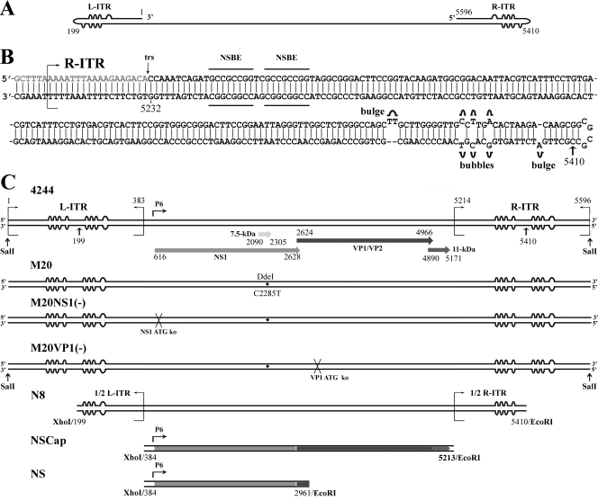
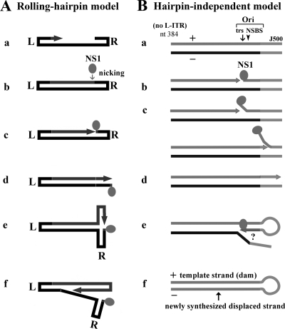
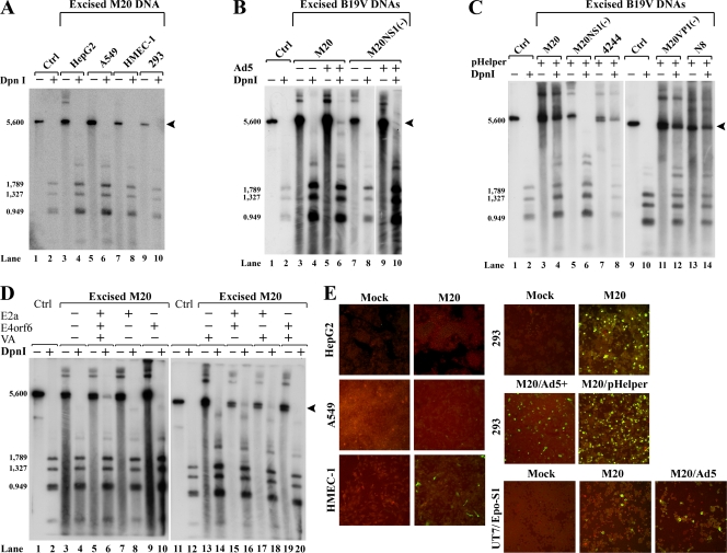
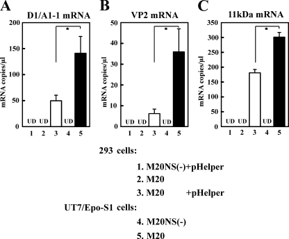
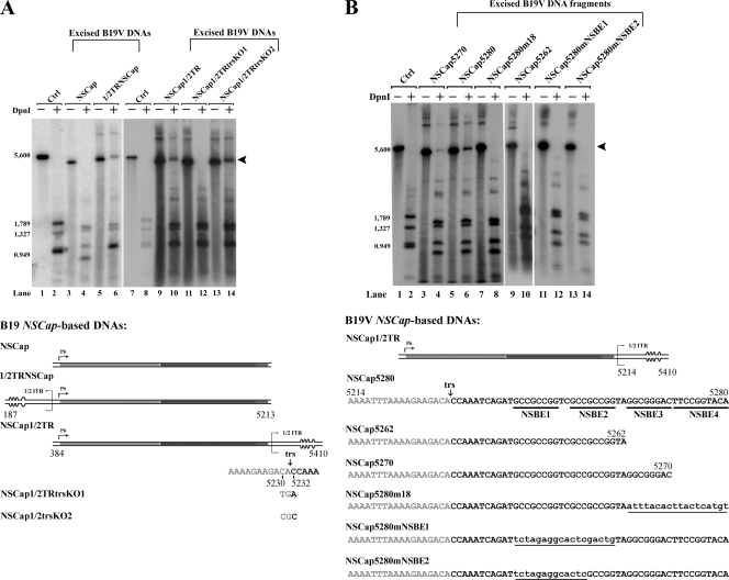
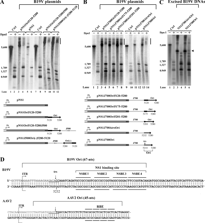
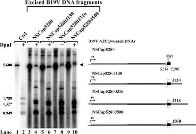
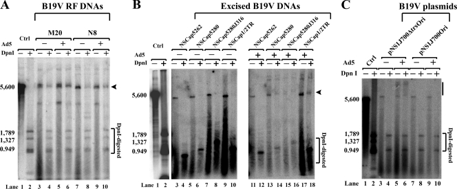
Human parvovirus B19 (B19V) is a member of the genus Erythrovirus in the family Parvoviridae. In vitro, autonomous B19V replication is limited to human erythroid progenitor cells and in a small number of erythropoietin-dependent human megakaryoblastoid and erythroid leukemic cell lines. Here we report that the failure of B19V DNA replication in nonpermissive 293 cells can be overcome by adenovirus infection. More specifically, the replication of B19V DNA in the 293 cells and the production of infectious progeny virus were made possible by the presence of the adenovirus E2a, E4orf6, and VA RNA genes that emerged during the transfection of the pHelper plasmid. Using this replication system, we identified the terminal resolution site and the nonstructural protein 1 (NS1) binding site on the right terminal palindrome of the viral genome, which is composed of a minimal origin of replication spanning 67 nucleotides. Plasmids or DNA fragments containing an NS1 expression cassette and this minimal origin were able to replicate in both pHelper-transfected 293 cells and B19V-semipermissive UT7/Epo-S1 cells. Our results have important implications for our understanding of native B19V infection.


Figures

FIG. 1.
FIG. 1.
Structure of the B19V genome. (A) Schematic diagram of the minus strand of the B19V genome. Identical ITRs are present at each end of the genome, and these are depicted with unpaired or mismatched bases in the palindromes represented by “bulges” or “bubbles,” respectively. (B) Structure of the B19V R-ITR. The palindrome at 365 nt is depicted in the “flip” orientation, and the putative trs and NS1 binding elements (NSBE) thought to comprise the NS1 binding site are indicated. Nucleotide numbers are according to GenBank accession no. AY386330. The ITR is an almost-perfect duplex structure; the only exceptions are the three unpaired bases at two sites (bulges) and three mismatched bases at three sites (bubbles). (C) Schematic diagrams of B19V DNAs. B19V DNA 4244 is the duplex RF of the B19V genome, which has the capability to replicate and produce progeny virus in B19V-permissive cells (54). The bubbles within each ITR reflect potential interstrand folding. The position of the P6 promoter, as well as the ORFs for the five B19V proteins, is also indicated. B19V DNA M20 is likewise an infectious DNA and is nearly identical to 4244 RF DNA; it contains the single nucleotide substitution C2285T to distinguish it from the native 4244 RF DNA by the digestion of DdeI as shown (54). In B19V DNAs M20NS(−) and M20VP1(−), the start codons for NS1 and VP1 are knocked out, respectively. In B19V DNA N8, only half of the L-ITR (nt 199 to 383) and half of the R-ITR (nt 5214 to 5410) sequences are included. The palindromic structure is not present in N8, and the bubbles, which are shown only as a symbol of half of the ITR, will thus not be folded in strand. The B19V DNAs of NSCap and NS do not contain any ITR sequences and encode different subsets of the B19V proteins. The ORFs preserved in the NSCap and NS DNAs are depicted in the respective diagrams, and the nucleotide numbers of each DNA are indicated beneath the diagram. The restriction enzymes to excise these B19V DNAs from the corresponding B19V plasmids described in Materials and Methods are indicated at their ends.
FIG. 2.
FIG. 2.
Models of parvovirus DNA replication. (A) Rolling-hairpin model of Dependovirus replication (13, 45). (a) The single-stranded DNA genome uses the hairpin as a primer for the synthesis of a duplex DNA molecule that is covalently closed at one end by the hairpin structure. (b) The ITR is cleaved at the nicking site by the NS1 protein (filled circle). (c, d) The hairpinned ITR is subsequently repaired, and this results in an open-ended duplex replication intermediate. (e, f) The repaired ITR is then denatured and reannealed, in a process termed reinitiation, to form a double-hairpinned intermediate that initiates a round of strand displacement synthesis. L, L-ITR; R, R-ITR. (B) Putative model of hairpin-independent B19V DNA replication in the current study. (a) A representative NSCap-based DNA fragment that contains the Ori, and a 500-nt extension, is shown, with each strand labeled as positive (+) or negative (−); no hairpin of more than 3 bp is present at the right end. (b) The Ori at the right end is nicked at the trs by NS1 (filled circle), which binds to the NS1 binding site (NSBS). (c) DNA is synthesized and primed from the free nucleotide (3′ OH) that is created by the nicking, with the complementary strand (negative [−]) serving as the template. (d) DNA synthesis goes to the 3′ end. (e) Following complementary-strand synthesis, the 3′ primer (possibly the last synthesized nucleotide) is used to initiate strand-displacement synthesis, through an unknown mechanism in which NS1 likely plays a key role. (f) The final replicated DNA product is composed of the prokaryotic template strand (+) and the newly synthesized complementary strand (−) which is resistant to DpnI digestion. dam, dam methylation.
FIG. 3.
FIG. 3.
Adenovirus infection and transfection with adenovirus gene products support B19V DNA replication in 293 cells. (A to D) Various cell types were transfected with excised B19V DNAs. Hirt DNA samples were extracted either from isolated nuclei (A) or from intact cells (B to D) and were used to test for DNA replication by Southern blotting using the B19V NSCap probe, with DpnI digestion (DpnI+) versus EcoRI digestion (DpnI−), revealing the levels of newly replicated and total input B19V DNA, respectively. In each experiment, DpnI− and DpnI+ M20 DNA (6 ng) were run in lanes 1 and 2, respectively, to serve as controls for DpnI digestion and as DNA size markers. Lanes 9 and 10 in panel C and lanes 11 and 12 in panel D are the same controls but for the respective blots. Arrowheads indicate DpnI-resistant DNA bands in DpnI-digested samples. (A) Infectious M20 DNA was transfected into HepG2, A549, HMEC-1, and 293 cells. (B) Both M20 and noninfectious M20NS1(−) DNA were transfected into 293 cells with or without Ad5 infection (Ad5+ or Ad5−). (C) A variety of infectious B19V DNAs [M20, M20VP1(−), or 4244] or noninfectious B19V DNAs [M20NS1(−) and N8] were transfected into 293 cells, either alone (pHelper−) or with pHelper (pHelper+), as indicated. (D) M20 was transfected into 293 cells along with different combinations of three adenovirus genes (E2a, E4orf6, and VA RNA) as indicated. (E) M20 DNA was transfected into HepG2, A549, HMEC-1, 293, or UT7/Epo-S1 cells, which were subsequently examined for NS1 expression. In the case of the 293 cells, cotransfection with pHelper was also tested, as was transfection into cells infected with Ad5 (Ad5+), as indicated. UT7/Epo-S1 cells also were tested in the presence or absence of Ad5 infection as indicated. NS1 expression was detected by immunofluorescence, using a polyclonal antibody against NS1 and a fluorescein isothiocyanate-conjugated secondary antibody (green); the cells were counterstained with Evans blue (red). The confocal images were taken at ×20 magnification with an Eclipse C1 Plus confocal microscope (Nikon). Ctrl, control.
FIG. 4.
FIG. 4.
Cotransfection of B19V RF M20 DNA into 293 cells with pHelper generates B19V progeny virus at a level comparable to that produced in UT7/Epo-S1 cells. 293 and UT7/Epo-S1 cells transfected with B19V DNAs were tested for the efficiency of virus production, based on the ability of lysates to stimulate virus mRNA production in CD36+ EPCs. Transfections were carried out on 2 × 10⁶ cells per sample, using B19V infectious RF M20 DNA or noninfectious M20NS(−) DNA; transfections in 293 cells were carried out with or without the cotransfection of pHelper. Cell lysates prepared at 3 days posttransfection were used to infect CD36+ EPCs, and the infectivity of these cell lysates was quantified by a real-time RT-PCR strategy that detected specifically D1/A1-1 (A), VP2 (B), and 11-kDa-encoding mRNAs (C) in the CD36+ EPC lysates. In each sample, the absolute number of mRNA copies was normalized to the level of β-actin mRNA (104 copies per μl). Results shown represent the averages and standard deviations of data from at least three independent experiments. UD stands for undetectable. A single star indicates a P value of <0.05, as assessed based on the Student's t test.
FIG. 5.
FIG. 5.
Identification of the trs and the NS1 binding site within the B19V R-ITR. Hirt DNA samples were extracted from intact cells. Southern blotting and testing for DNA replication, as described in the legend to Fig. 3, were carried out on additional excised B19V DNA fragments. (A) NSCap and four derivatives were tested to identify the trs. In one derivative, half of the L-ITR (1/2TRNSCap) is included; in a second, half of the R-ITR (NSCap1/2TR) is included. In both the third and fourth NSCap1/2TR-derived constructs, nucleotides of the putative trs are mutated. (B) Six additional mutants affecting nucleotides in the R-ITR were tested to identify the NS1 binding elements (NSBE) of the NS1 binding site. Three constructs are simply truncations. In the others, which harbor mutations in addition to being truncated at nt 5280, the mutated nucleotides are indicated as small characters and underlined.
FIG. 6.
FIG. 6.
Identification of the B19V Ori. Hirt DNA samples were extracted from intact cells. Southern blotting and testing for DNA replication, as described in the legend to Fig. 3, was carried out on B19V DNA plasmids (A, B) and excised B19V DNA fragments (C). (A, B) The B19V plasmids schematically depicted at the bottoms of the panels were tested. The locations, positions, and lengths of the putative Ori are indicated, as are the lengths and orientations of the prokaryotic sequences that were inserted as described in Materials and Methods. Bars indicate smears of the DpnI-resistant DNA band in DpnI-digested samples. (C) Two B19V DNA fragments were excised from plasmids pNS1J700Ori and pNS1J700ΔtrsOri through XhoI/EcoRI digestion, respectively, and tested for DpnI-resistant DNA. The arrowhead indicates the DpnI-resistant DNA band in DpnI-digested samples. (D) Structure comparison of the Ori between B19V and AAV2. The nucleotide sequences of the region spanning the B19V and AAV2 Ori, as indicated, are shown, and the identified trs and NS1 binding elements (NSBE) that comprise the NS1 binding site are indicated. Ctrl, control; RBE, Rep binding element.
FIG. 7.
FIG. 7.
Hairpin-independent replication of B19V DNA. Hirt DNA samples were extracted from intact cells. Southern blotting and testing for DNA replication, as described in the legend to Fig. 3, were carried out on excised B19V NSCap5280 DNA and three variants, in which heterologous prokaryotic fragments of various lengths were fused to the parent DNA, as illustrated to the right. The arrowhead indicates the DpnI-resistant DNA band in DpnI-digested samples.
FIG. 8.
FIG. 8.
Adenovirus facilitates B19V DNA replication in UT7/Epo-S1 cells. Hirt DNA samples were extracted from intact cells. Southern blotting and testing for DNA replication, as described in the legend to Fig. 3, were carried out on excised B19V DNA fragments (A, B) and B19V DNA plasmids (C) transfected into the B19V-semipermissive UT7/Epo-S1 cells. As indicated, six B19V DNAs (A, B) and two B19V plasmids (C) were tested in either Ad5-infected (+) or uninfected (−) cells. Arrowheads in panels A and B and the bar in panel C identify the DpnI-resistant DNA band in DpnI-digested samples.

Similar articles

Cited by

References

    1. Anderson, M. J., P. G. Higgins, L. R. Davis, J. S. Willman, S. E. Jones, I. M. Kidd, J. R. Pattison, and D. A. Tyrrell. 1985. Experimental parvoviral infection in humans. J. Infect. Dis. 152:257-265. - PubMed
    1. Astell, C. R., W. Luo, J. Brunstein, and J. Amand. 1997. B19 parvovirus: biochemical and molecular features, p. 16-41. In L. J. Anderson and N. Young (ed.), Human parvovirus B19. Karger, Basel, Switzerland.
    1. Berns, K. I., and C. R. Parrish. 2007. Parvoviridae, p. 2437-2477. In D. M. Knipe and P. M. Howley (ed.), Fields virology, 5th ed. Lippincott Williams, New York, NY.
    1. Bloom, M. E., and N. Young. 2001. Parvoviruses, p. 2361-2379. In D. M. Knipe (ed.), Fields virology. Lippincott Williams and Wilkins, Philadelphia, PA.
    1. Brown, K. E., and N. Young. 1997. Human parvovirus B19: pathogenesis of disease, p. 105-119. In L. J. Anderson and N. Young (ed.), Human parvovirus B19, vol. 20. Karger, Basel, Switzerland.

Publication types