. 2009 Sep;83(18):9370-87.
doi: 10.1128/JVI.02076-08. Epub 2009 Jul 8.

Insight into poliovirus genome replication and encapsidation obtained from studies of 3B-3C cleavage site mutants

Affiliations

Insight into poliovirus genome replication and encapsidation obtained from studies of 3B-3C cleavage site mutants

Hyung Suk Oh et al. J Virol. 2009 Sep.

Abstract

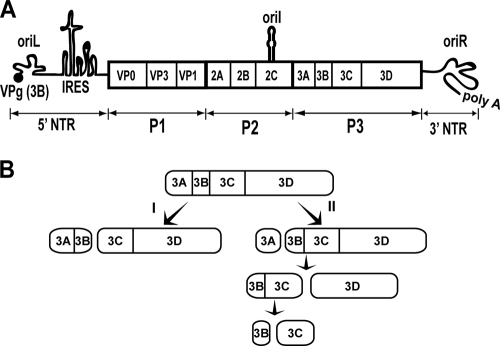
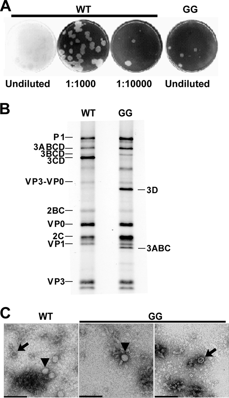
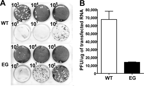
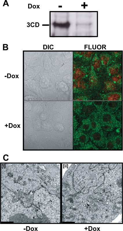
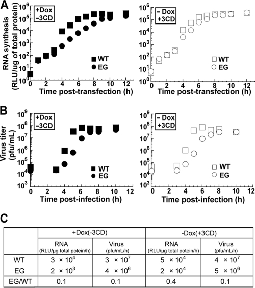
A poliovirus (PV) mutant (termed GG), which is incapable of producing 3AB, VPg, and 3CD proteins due to a defective cleavage site between the 3B and 3C proteins, replicated, producing 3BC-linked RNA rather than the VPg-linked RNA produced by the wild type (WT). GG PV RNA is quasi-infectious. The yield of infectious GG PV relative to replicated RNA is reduced by almost 5 logs relative to that of WT PV. Proteolytic activity required for polyprotein processing is normal for the GG mutant. 3BC-linked RNA can be encapsidated as efficiently as VPg-linked RNA. However, a step after genome replication but preceding virus assembly that is dependent on 3CD and/or 3AB proteins limits production of infectious GG PV. This step may involve release of replicated genomes from replication complexes. A pseudorevertant (termed EG) partially restored cleavage at the 3B-3C cleavage site. The reduced rate of formation of 3AB and 3CD caused corresponding reductions in the observed rate of genome replication and infectious virus production by EG PV without impacting the final yield of replicated RNA or infectious virus relative to that of WT PV. Using EG PV, we showed that genome replication and encapsidation were distinct steps in the multiplication cycle. Ectopic expression of 3CD protein reversed the genome replication phenotype without alleviating the infectious-virus production phenotype. This is the first report of a trans-complementable function for 3CD for any picornavirus. This observation supports an interaction between 3CD protein and viral and/or host factors that is critical for genome replication, perhaps formation of replication complexes.


Figures

FIG. 1.
FIG. 1.
PV genome organization and P3 processing pathway. (A) Schematic of the PV genome. The 5′ end of the genome is covalently linked to a peptide (VPg) encoded by the 3B region of the genome. The 3′ end contains a poly(rA) tail. Three cis-acting replication elements are known. oriL is located in 5′ NTR. oriR is located in the 3′ NTR. oriI is located in 2C-coding sequence for PV; the position of this element is virus dependent. oriI is the template for VPg uridylylation. Translation initiation employs an internal ribosome entry site (IRES). The single open reading frame encodes a polyprotein. P1 produces virion structural proteins as indicated. P2 produces proteins thought to participate in virus-host interactions required for genome replication. P3 produces proteins thought to participate directly in genome replication. Polyprotein processing is mediated by protease activity residing in 2A, 3C, and/or 3CD proteins. (B) Processing of the P3 precursor occurs by two independent pathways (60). There are major (I) and minor (II) pathways. In pathway I, processing between 3B and 3C yields 3AB and 3CD. In pathway II, processing between 3A and 3B yields 3A and 3BCD. 3BCD processing yields 3BC and 3D; 3BC processing yields 3B and 3C. Pathway II is proposed to function in genome replication and is not perturbed in the GG mutant.
FIG. 2.
FIG. 2.
Changing the 3B-3C cleavage site from QG to GG produces a quasi-infectious virus. (A) Specific infectivity of GG mutant and WT PV RNA. HeLa cells were transfected with GG mutant or WT PV RNA, diluted, added to HeLa cell monolayers, overlaid with agarose, and held at 34°C for 5 days, at which time the agarose overlay was removed and cells were stained with crystal violet. From each of the five plaques obtained for the GG mutant RNA transfection, virus was plaque purified and viral RNA was extracted, reverse transcribed, and sequenced. Sequencing identified a single mutation at the 3B-3C cleavage site that converted GG to EG. (B) Processing evaluated by cell-free translation. HeLa cell-free translation extracts containing [35S]methionine and [35S]cysteine were programmed with WT or GG mutant RNA. Radiolabeled proteins were separated by 15% SDS-PAGE and detected by phosphorimaging. The bands corresponding to the different precursor and processed proteins expected for the WT are indicated on the left. The identities of bands unique to the GG mutant are indicated on the right. (C) TEM of WT and GG virus particles. HeLa cells were transfected with GG mutant or WT PV RNA and incubated at 34°C for 20 h, at which time cells were harvested and lysed with three freeze-thaw cycles. WT and GG mutant PV particles were then purified by precipitation using 10% PEG 8000 followed by centrifugation through a 30% sucrose cushion. Purified virus particles were negatively stained with 2% UA and observed by TEM. Bars = 0.1 μm. Representative virus particles that are empty (arrows) or contain packaged RNA (▾) are indicated.
FIG. 3.
FIG. 3.
Changing the 3B-3C cleavage site from GG to EG restores infectivity. (A) Infectious center assay for EG mutant and WT PV RNA. HeLa cells were transfected with in vitro-transcribed EG mutant or WT PV RNA, diluted, added to HeLa cell monolayers, overlaid with agarose, and held at 37°C for 2 days, at which time the agarose overlay was removed and cells were stained with crystal violet. (B) Quantitation of data shown in panel A. The specific infectivities for WT and EG mutant RNA are 68,000 ± 10,000 and 14,000 ± 1,000 PFU/μg RNA, respectively.
FIG. 4.
FIG. 4.
EG PV exhibits a decreased rate of RNA synthesis and virus production. (A) Kinetics of virus production by WT and EG PV. HeLa cells were infected with WT or EG PV (MOI, 10) and held at 37°C. At the indicated times postinfection, cells were harvested and lysed by freeze-thawing and virus was titered. (B) Kinetics of RNA synthesis by WT, EG mutant, and Y3F mutant subgenomic replicon RNA. The Y3F replicon encodes a 3B with a change of tyrosine-3 to phenylalanine. Y-3 is the nucleophile employed to form VPg-pU. Products formed in this background would be irrelevant. HeLa cells were transfected with in vitro-transcribed replicon RNA and held at 37°C, and luciferase activity was monitored for 12 h posttransfection. (C) Kinetics of translation by WT and EG mutant subgenomic replicon RNA in the presence of brefeldin A (BFA). HeLa cells were transfected with in vitro-transcribed replicon RNA, incubated in the presence of brefeldin A (2 μg/ml), and held at 37°C, and luciferase activity was monitored for 12 h posttransfection.
FIG. 5.
FIG. 5.
EG PV exhibits a post-genome replication, pre-virus assembly defect. (A) Experimental design. HeLa cells were infected with WT or EG PV (MOI, 1) in the presence of hydantoin and held at 37°C for 6 or 8 h, at which time the medium containing hydantoin was removed, cells were washed with PBS, fresh medium was added, and the infected cells were returned to 37°C. At the indicated times postinfection, cells were harvested and lysed by multiple freeze-thaw cycles and virus was titered. (B and C) Virus titer for WT and EG PV from the experiment described in panel A when hydantoin was removed at 6 (B) or 8 (C) h postinfection. (D) TEM of WT and EG virus particles. WT and EG PV particles were purified by precipitation using 10% PEG 8000 followed by centrifugation through a 30% sucrose cushion. Purified virus particles were negatively stained with 2% UA and observed by TEM. Representative virus particles that are empty (arrow) or contain packaged RNA (▾) are indicated. Bars = 0.1 μm.
FIG. 6.
FIG. 6.
EG PV produced in the presence of hydantoin does not require new RNA synthesis. (A) Cordycepin blocks new RNA synthesis. HeLa cells were transfected with in vitro-transcribed WT subgenomic replicon RNA, incubated with cordycepin at the indicated concentrations (μM), and held at 37°C, and luciferase activity was monitored for 8 h posttransfection. (B) Cordycepin blocks ongoing RNA synthesis. HeLa cells were transfected with in vitro-transcribed WT subgenomic replicon RNA and held at 37°C. At 0 or 3 h posttransfection, 200 μM cordycepin was added and luciferase activity was monitored. A control experiment was performed in the absence of cordycepin. (C) The hydantoin experiment in the presence and absence of cordycepin. HeLa cells were infected with WT or EG PV (MOI, 1) in the presence of hydantoin and held at 37°C for 6 or 8 h, at which time either 0 or 300 μM cordycepin was added. At 6.5 or 8.5 h, the medium was removed, cells were washed with PBS, fresh medium containing either no or 300 μM cordycepin was added, and the cells were returned to 37°C. At the indicated times postinfection, cells were harvested, lysed by freeze-thawing, and virus titered. (D) Kinetics of WT PV production after removal of hydantoin (Hyd) is not altered by the cordycepin block. The experiment was performed as described for panel C. Data shown were obtained in the presence or absence of cordycepin (Cor). (E) Kinetics of EG PV production after removal of hydantoin is not altered by the cordycepin block. The experiment was performed as described for panel C. Data shown were obtained in the presence or absence of cordycepin.
FIG. 7.
FIG. 7.
EG PV exhibits delayed kinetics of P3 processing. (A) EG restores P3 processing. Processing was evaluated by Western blotting. HeLa cells were transfected with WT, GG, or EG subgenomic replicon RNA, held at 34°C, and harvested 20 h posttransfection. Extracts were prepared and processed for Western blotting as described in Materials and Methods. Antisera against VPg, 3C, and 3D were employed. The bands corresponding to the different precursor and processed proteins are indicated. (B) Kinetics of P3 processing for EG is delayed. Processing was evaluated by Western blotting. HeLa cells were transfected with WT or EG mutant subgenomic replicon RNA, held at 34°C, and harvested at 6, 8, and 20 h posttransfection. Extracts were prepared and processed for Western blotting as described in Materials and Methods. Antisera against VPg and 3D were employed. The bands corresponding to the different precursor and processed proteins are indicated. Purified 3CD and 3D (M1) or 3ABC and 3BC (M2) were loaded in lanes 8 and 17 and 9 and 18 as positive controls. Uninfected cells were used as negative controls (lanes 1 and 10). (C) Processing was evaluated by cell-free translation. HeLa cell-free translation extracts containing [35S]methionine and [35S]cysteine were programmed with WT or EG mutant RNA for 0.25, 1, and 2 h. Radiolabeled proteins were separated by 15% SDS-PAGE and detected by phosphorimaging. The bands corresponding to the different precursor and processed proteins are indicated.
FIG. 8.
FIG. 8.
3B precursor-linked RNA is produced by EG PV and converted to VPg-linked RNA prior to the hydantoin-sensitive step. Interrogation of protein linkage to WT and EG mutant RNA was analyzed by RNA immunoprecipitation and Northern blotting. (A) RNA isolated from replicon RNA-transfected cells. HeLa cells were transfected with WT, EG, or Y3F RNA and held at 37°C for 10 h. Total RNA was isolated from transfected HeLa cells and immunoprecipitated using antibodies against VPg, 3C, 3D, or HCV NS5A (as a negative control). The immunoprecipitated RNA was separated on a 0.6% agarose gel containing 0.8 M formaldehyde, transferred to a nylon membrane, and hybridized with a 32P-labeled DNA probe. The hybridized DNA probe was visualized by phosphorimaging. Shown is a phosphorimage after a 1-day exposure. In vitro-transcribed RNA is shown as a reference. (B) RNA isolated at early times postinfection. HeLa cells were infected with WT PV (MOI, 10) and held at 37°C for 0, 4, or 5 h. As a positive control, EG PV (MOI, 10)-infected HeLa cells were incubated at 37°C for 10 h. Total RNA was isolated from infected HeLa cells and immunoprecipitated using antibodies against VPg, 3C, or HCV NS5A. The immunoprecipitated RNA was separated as described above. In vitro-transcribed RNA is shown as a reference. (C) RNA isolated from HeLa cells infected with WT PV or EG PV (MOI, 10) in the presence (+Hyd) or absence (−Hyd) of hydantoin at 37°C for 6 h (WT PV) or 8 h (EG PV). Total RNA was immunoprecipitated using antibodies against VPg and 3C. The immunoprecipitated RNA was separated as described above. WT or EG PV RNA used for immunoprecipitation is shown in lanes 1 to 4. (D) RNA isolated from purified virus particles. HeLa cells were infected with WT or EG PV and held at 37°C until CPE. Cells were harvested, and WT and EG PV was purified by precipitation using 8 to 10% PEG 8000 followed by centrifugation through a 30% sucrose cushion. Viral RNA was isolated from purified viruses and immunoprecipitated using antibodies against VPg, 3C, 3D, or HCV NS5A. The immunoprecipitated RNA was separated as described above. Total RNA purified from Y3F mutant RNA-transfected cells was used as a negative control.
FIG. 9.
FIG. 9.
Establishment of a HeLa cell line capable of regulated expression of PV 3CD. HeLa-3CD cell lines were incubated in the absence (−) or presence (+) of Dox (1 μg/ml) for 36 h at 37°C as described in Materials and Methods. (A) Dox removal induces 3CD expression. 3CD induction was confirmed by Western blot of total cellular protein using anti-3D antisera. (B) Expressed 3CD localizes to the nucleus. Immunofluorescence of HeLa-3CD cell lines using anti-3D (red) and antiactin (green) labeled antibodies. (C) 3CD expression is not cytopathic. HeLa-3CD cells were fixed and visualized by electron microscopy. Bars = 2 μm. HeLa-3CD cells appear normal both in the absence (−) and presence (+) of Dox.
FIG. 10.
FIG. 10.
Ectopic expression of 3CD enhances the kinetics of RNA synthesis of EG PV without impacting the kinetics of virus production. (A) Kinetics of RNA synthesis by WT and EG mutant subgenomic replicon RNA in the absence (+Dox, −3CD) or presence (−Dox, +3CD) of ectopic expression of 3CD. HeLa-3CD cells incubated in the presence or absence of Dox were transfected with either WT or EG in vitro-transcribed replicon RNA and held at 37°C, and luciferase activity was monitored for 12 h posttransfection. The kinetics of RNA synthesis for EG expressing 3CD (○) are essentially identical to that of the WT (□). There was no difference in the kinetics of RNA synthesis for WT replicon in the presence or absence of 3CD. (B) Kinetics of virus production by WT and EG PV in the absence or presence of ectopic expression of 3CD. HeLa-3CD cells incubated in the presence or absence of Dox were infected with WT or EG PV (MOI, 10) and held at 37°C. At the indicated times postinfection, cells were harvested and lysed by freeze-thawing and virus was titered. There was no difference in the kinetics of EG mutant virus production in the absence (•) or presence (○) of 3CD. (C) Average rates of RNA synthesis (RNA) and infectious virus production (virus) in the absence or presence of ectopic expression of 3CD for WT and EG. Rates shown were calculated from data obtained between 3 and 6 h, inclusive, posttransfection or postinfection.
FIG. 11.
FIG. 11.
Implications of this study presented in the context of the PV multiplication cycle. Roles for P3 proteins suggested by this study appear in boxes, and the additional step appears as a thick arrow. Genomic RNA is first translated to produce the PV polyprotein. Translation is inhibited by cycloheximide. The polyprotein is co- and posttranslationally processed to produce the various precursors and processed proteins that are needed for PV multiplication. This study suggests that 3ABCD and/or 3BCD efficiently cleaves the PV polyprotein. RCs are formed, followed by genome replication. This study implicates 3CD in a step prior to genome replication. We have assigned this function of 3CD to RC formation. However, additional studies will be required to confirm this possibility. Early during infection, replicated RNA enters the pool of transfected RNA. This study implicates 3AB or 3CD in a step after genome replication but prior to virus assembly. We propose that this step permits transit of replicated RNA from RCs to virus particles. Finally, virus assembly, i.e., encapsidation and maturation, occurs. Hydantoin can inhibit production of infectious virus at the virus assembly step.

Similar articles

Cited by

References

    1. Agol, V. I., A. V. Paul, and E. Wimmer. 1999. Paradoxes of the replication of picornaviral genomes. Virus Res. 62:129-147. - PubMed
    1. Aldabe, R., and L. Carrasco. 1995. Induction of membrane proliferation by poliovirus proteins 2C and 2BC. Biochem. Biophys. Res. Commun. 206:64-76. - PubMed
    1. Amineva, S. P., A. G. Aminev, A. C. Palmenberg, and J. E. Gern. 2004. Rhinovirus 3C protease precursors 3CD and 3CD′ localize to the nuclei of infected cells. J. Gen. Virol. 85:2969-2979. - PubMed
    1. Andino, R., G. E. Rieckhof, P. L. Achacoso, and D. Baltimore. 1993. Poliovirus RNA synthesis utilizes an RNP complex formed around the 5′-end of viral RNA. EMBO J. 12:3587-3598. - PMC - PubMed
    1. Andino, R., G. E. Rieckhof, and D. Baltimore. 1990. A functional ribonucleoprotein complex forms around the 5′ end of poliovirus RNA. Cell 63:369-380. - PubMed

Publication types

LinkOut - more resources