

Transcriptional changes in the brains of cattle orally infected with the bovine spongiform encephalopathy agent precede detection of infectivity
- PMID: 19587050
- PMCID: PMC2738270
- DOI: 10.1128/JVI.00352-09
Transcriptional changes in the brains of cattle orally infected with the bovine spongiform encephalopathy agent precede detection of infectivity
Abstract
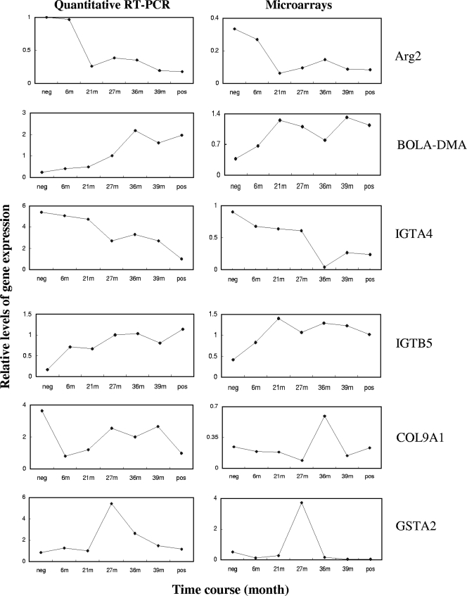
Bovine spongiform encephalopathy (BSE) is a fatal, transmissible, neurodegenerative disease of cattle. BSE can be transmitted experimentally between cattle through the oral route, and in this study, brain tissue samples from animals at different time points postinoculation were analyzed for changes in gene expression. The aims of this study were to identify differentially regulated genes during the progression of BSE using microarray-based gene expression profiling and to understand the effect of prion pathogenesis on gene expression. A total of 114 genes were found to be differentially regulated over the time course of the infection, and many of these 114 genes encode proteins involved in immune response, apoptosis, cell adhesion, stress response, and transcription. This study also revealed a broad correlation between gene expression profiles and the progression of BSE in cattle. At 21 months postinoculation, the largest number of differentially regulated genes was detected, suggesting that there are many pathogenic processes in the animal brain even prior to the detection of infectivity in the central nervous systems of these orally infected cattle. Moreover, evidence is presented to suggest that it is possible to predict the infectious status of animals using the expression profiles from this study.
Figures
Similar articles
-
Whole Blood Gene Expression Profiling in Preclinical and Clinical Cattle Infected with Atypical Bovine Spongiform Encephalopathy.PLoS One. 2016 Apr 13;11(4):e0153425. doi: 10.1371/journal.pone.0153425. eCollection 2016. PLoS One. 2016. PMID: 27073865 Free PMC article.
-
Transcriptional analysis implicates endoplasmic reticulum stress in bovine spongiform encephalopathy.PLoS One. 2010 Dec 3;5(12):e14207. doi: 10.1371/journal.pone.0014207. PLoS One. 2010. PMID: 21151970 Free PMC article.
-
Microarray analysis in caudal medulla of cattle orally challenged with bovine spongiform encephalopathy.Genet Mol Res. 2011 Oct 25;10(4):3948-62. doi: 10.4238/2011.October.25.5. Genet Mol Res. 2011. PMID: 22033911
-
The research programme on transmissible spongiform encephalopathies in Britain with special reference to bovine spongiform encephalopathy.Dev Biol Stand. 1993;80:157-70. Dev Biol Stand. 1993. PMID: 8270105 Review.
-
Prion biology relevant to bovine spongiform encephalopathy.J Anim Sci. 2005 Jun;83(6):1455-76. doi: 10.2527/2005.8361455x. J Anim Sci. 2005. PMID: 15890824 Review.
Cited by
-
Proteomic consequences of expression and pathological conversion of the prion protein in inducible neuroblastoma N2a cells.Prion. 2010 Oct-Dec;4(4):292-301. doi: 10.4161/pri.4.4.13435. Epub 2010 Oct 27. Prion. 2010. PMID: 20930564 Free PMC article.
-
Central nervous system gene expression changes in a transgenic mouse model for bovine spongiform encephalopathy.Vet Res. 2011 Oct 28;42(1):109. doi: 10.1186/1297-9716-42-109. Vet Res. 2011. PMID: 22035425 Free PMC article.
-
RNA editing alterations define manifestation of prion diseases.Proc Natl Acad Sci U S A. 2019 Sep 24;116(39):19727-19735. doi: 10.1073/pnas.1803521116. Epub 2019 Sep 6. Proc Natl Acad Sci U S A. 2019. PMID: 31492812 Free PMC article.
-
Functional genomics approach for identification of molecular processes underlying neurodegenerative disorders in prion diseases.Curr Genomics. 2012 Aug;13(5):369-78. doi: 10.2174/138920212801619223. Curr Genomics. 2012. PMID: 23372423 Free PMC article.
-
Whole Blood Gene Expression Profiling in Preclinical and Clinical Cattle Infected with Atypical Bovine Spongiform Encephalopathy.PLoS One. 2016 Apr 13;11(4):e0153425. doi: 10.1371/journal.pone.0153425. eCollection 2016. PLoS One. 2016. PMID: 27073865 Free PMC article.
References
-
- Affymetrix UK Ltd. 2001. GeneChip expression analysis technical manual. Affymetrix, Ltd., Santa Clara, CA. http://www.affymetrix.com/support/downloads/manuals/expression_analysis_....
-
- Aguzzi, A. 2006. Prion diseases of humans and farm animals: epidemiology, genetics, and pathogenesis. J. Neurochem. 97:1726-1739. - PubMed
-
- Aguzzi, A., C. Sigurdson, and M. Heikenwalder. 2008. Molecular mechanisms of prion pathogenesis. Annu. Rev. Pathol. 3:11-40. - PubMed
-
- Arnold, M. E., J. B. Ryan, T. Konold, M. M. Simmons, Y. I. Spencer, A. Wear, M. Chaplin, M. Stack, S. Czub, R. Mueller, P. R. Webb, A. Davis, J. Spiropoulos, J. Holdaway, S. A. Hawkins, A. R. Austin, and G. A. Wells. 2007. Estimating the temporal relationship between PrPSc detection and incubation period in experimental bovine spongiform encephalopathy of cattle. J. Gen. Virol. 88:3198-3208. - PubMed
Publication types
MeSH terms
LinkOut - more resources
Full Text Sources
