. 2009 Sep;83(18):9131-9.
doi: 10.1128/JVI.00871-09. Epub 2009 Jul 8.

Two small RNAs encoded within the first 1.5 kilobases of the herpes simplex virus type 1 latency-associated transcript can inhibit productive infection and cooperate to inhibit apoptosis

Affiliations

Two small RNAs encoded within the first 1.5 kilobases of the herpes simplex virus type 1 latency-associated transcript can inhibit productive infection and cooperate to inhibit apoptosis

Wenwen Shen et al. J Virol. 2009 Sep.

Abstract

The herpes simplex virus type 1 (HSV-1) latency-associated transcript (LAT) is abundantly expressed in latently infected trigeminal ganglionic sensory neurons. Expression of the first 1.5 kb of LAT coding sequences is sufficient for the wild-type reactivation phenotype in small animal models of infection. The ability of the first 1.5 kb of LAT coding sequences to inhibit apoptosis is important for the latency-reactivation cycle. Several studies have also concluded that LAT inhibits productive infection. To date, a functional LAT protein has not been identified, suggesting that LAT is a regulatory RNA. Two small RNAs (sRNAs) were previously identified within the first 1.5 kb of LAT coding sequences. In this study, we demonstrated that both LAT sRNAs were expressed in the trigeminal ganglia of mice latently infected with an HSV-1 strain that expresses LAT but not when mice were infected with a LAT null mutant. LAT sRNA1 and sRNA2 cooperated to inhibit cold shock-induced apoptosis in mouse neuroblastoma cells. LAT sRNA1, but not LAT sRNA2, inhibited apoptosis less efficiently than both sRNAs. When rabbit skin cells were cotransfected with plasmids that express LAT sRNA1 and HSV-1 genomic DNA, the amount of infectious virus released was reduced approximately 3 logs. Although LAT sRNA2 was less effective at inhibiting virus production, it inhibited expression of infected cell protein 4 (ICP4). Neither LAT sRNA had an obvious effect on ICP0 expression. These studies suggested that expression of two LAT sRNAs plays a role in the latency-reactivation cycle by inhibiting apoptosis and productive infection.


Figures

FIG. 1.
FIG. 1.
Schematic of HSV-1 and organization of the LAT locus. (A) The LAT locus is present in the unique long (UL) and unique short (US) regions. The primary 8.3-kb LAT is shown as a long arrow. The stable 2-kb LAT is shown as a solid rectangle. The LAT TATA box is denoted by TATA, and the small arrow and +1 indicate the start of LAT transcription (genomic nt 118801). The LAT promoter (LAP) is denoted by the gray rectangle. The relative locations of mRNAs encoding ICP0 and ICP34.5 are shown for reference. Expression of the first 1.5 kb of LAT coding sequences (+1,500) is sufficient for reactivation from latency (56). The relative locations of the two LAT sRNAs that were previously identified are denoted by asterisks (51). (B) Nucleotide sequences of the LAT sRNAs (51). The underlined ATGs correspond to the initiating ATGs of ORF4 (LAT sRNA1) or ORF8 (LAT sRNA2) (17). (C) Female Swiss Webster mice were infected with dLAT2903R (lane R) or dLAT2903 (lane 2903). At 30 days after infection, TG were extracted and sRNA was prepared by using the miRNA isolation kit (miVana; Ambion) according to the manufacturer's instructions. RT-PCR using adaptor primers and LAT-specific primers was used to examine sRNA expression, as described in Materials and Methods. (D) Double-stranded DNA corresponding to the respective sRNAs was synthesized and cloned into the siRNA expression plasmid, pSilencer 2.1-U6 neo (Ambion). neuro-2a cells were transfected with 5 μg of a plasmid expressing LAT sRNA1 (lane 1), LAT sRNA2 (lane 2), or the empty pSilencer 2.1-U6 neo (lane E). At 40 h after transfection, sRNA was prepared using the mirVana miRNA isolation kit (Ambion) according to the manufacturer's instructions, and the expression of LAT sRNA was examined by RT-PCR. Following purification of sRNA by the mirVana kit, total sRNA (1 μg) was electrophoresed on a 2% agarose gel to confirm that similar levels of RNA were used for RT-PCR amplification.
FIG. 2.
FIG. 2.
LAT sRNA2 inhibits ICP4 protein expression. Western blot analysis of ICP4 protein expression in RS cells (A), ICP4 protein expression in neuro2-A cells (B), or ICP0 expression in RS cells (D) was performed using commercially available antiserum, as described in Materials and Methods. Lane U, untransfected cells. Except for the U lane, all cells were transfected with 2 μg of the PN11 plasmid expressing ICP4 (A to C) or the ICP0 expression plasmid (2 μg plasmid) (D). Cultures were cotransfected with the following plasmids: 4 μg of the empty siRNA vector (lane E), 4 μg of plasmid expressing the negative control siRNA (lane NC), 4 μg of the plasmid expressing LAT sRNA1 (lane 1), 4 μg of the plasmid expressing LAT sRNA2 (lane 2), 2 μg of each plasmid expressing LAT sRNA1 and LAT sRNA2 (lane B), 4 μg of plasmid expressing mutant LAT sRNA1 (lane 1 M), 4 μg of plasmid expressing LAT sRNA1 (lane 2 M), and 2 μg of each plasmid expressing mutant LAT sRNA1 and LAT sRNA2 (lane BM). β-Actin was used as a protein loading control (bottom). A total of 350 μg of protein was loaded into each lane. As a positive control, RS cells were infected with dLAT2903 for 8 h, and total cell lysate was loaded (lane +). For panel C, total RNA was prepared and RT-PCR was performed using random primers. Amplification of ICP4 or GAPDH was performed using the primers described in Materials and Methods. Lane NT was a negative control that lacked template.
FIG. 3.
FIG. 3.
LAT sRNA1 and sRNA2 inhibit productive infection in RS cells. RS cells were transfected with HSV-1 DNA (2 μg of dLAT2903) and the designated plasmids that express LAT sRNAs. (A) Virus was collected from transfected cells at 48 h after transfection, and plaque assays were performed in RS cells. Bar NC, 4 μg of a plasmid expressing the control siRNA; bar E, 4 μg of the empty siRNA vector; bar 1, cells cotransfected with 4 μg of the plasmid that expresses LAT sRNA1; bar 2, 4 μg of the plasmid expressing LAT sRNA2; bar B, 2 μg of each plasmid expressing LAT sRNA1 and LAT sRNA2; bar 1M, 4 μg of the plasmid expressing mutant LAT sRNA1; bar 2M, 4 μg of the plasmid expressing mutant LAT sRNA2; and bar BM, 2 μg of each plasmid expressing mutant LAT sRNA1 and LAT sRNA2. The values are the averages of five independent experiments. Statistical analysis of the values was calculated using a P value unpaired test. (B) The designated samples were collected at 48 h after transfection, and Western blotting was performed using 600 μg protein for ICP4 and β-actin. Lane U, cells not transfected with any plasmid or viral DNA; lane E, empty siRNA vector; lane 1, cells cotransfected with 4 μg of the plasmid that expresses LAT sRNA1; lane 2, plasmid expressing LAT sRNA2; and lane +, cells infected with dLAT2903 (1 PFU/cell) for 8 h.
FIG. 4.
FIG. 4.
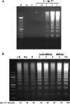
Analysis of cold shock-induced apoptosis in neuro-2A cells. (A) neuro-2A cells were subjected to cold shock-induced apoptosis, as described in Materials and Methods. Apoptotic DNA was isolated from 3 × 10⁵ cells/dish and electrophoresed in a 2% agarose gel. DNA was visualized after staining with EtBr and then photographed. Lane 2A represents actively growing neuro-2A cells and the 4° lane represents neuro-2A cells incubated at 4°C for 1 h. Cells were subjected to cold shock followed by incubation at 37°C for 1, 1.5, 2, 3, and 4 h, respectively, to induce apoptosis. The results are representative of more than 20 independent studies. (B) neuro-2A cells were transfected with pcDNA3.1 that expresses the BHV-1 LR gene (6 μg plasmid) (lane LR), the Bcl-2 gene (6 μg plasmid) (lane Bcl), plasmids expressing both LAT siRNAs (3 μg of each plasmid) (lane B), increasing concentrations of the siRNA expression plasmid containing LAT sRNA1, or the plasmid expressing LAT sRNA2 (6 μg plasmid DNA) (lane 6). neuro-2A cells transfected with pSilencer 2.1-U6 neo expressing the control siRNA were used as a negative control (6 μg plasmid) (lane NC). To maintain equal amounts of plasmid DNA, certain cultures were cotransfected with the empty siRNA expression vector. The relative amount (Rel.) of apoptotic DNA in the respective lanes from panel B was measured using Bio-Rad Molecular Imager FX. The results are representative of at least five independent experiments.
FIG. 5.
FIG. 5.
LAT sRNAs inhibit cell death. neuro-2A cells (3 × 10⁵) were transfected with the designated plasmids. (A) Following cold shock-induced apoptosis, nuclear DNA was stained with propidium iodide (50 μg/ml) and fluorescence-activated cell sorter analysis was performed to measure the sub-G1 levels of DNA. The values are the averages of five independent experiments. Column M, mock-transfected cells; bar NC, empty siRNA expression plasmid (6 μg DNA); bar 1, 3 μg of the plasmid expressing LAT sRNA1 and 3 μg of the empty siRNA expression plasmid; bar 2, 3 μg of the plasmid expressing LAT sRNA2 and 3 μg of the empty siRNA expression plasmid; bar B, 3 μg each of the LAT sRNA expression plasmids; and bar Bcl-2, 3 μg of a Bcl-2 expression plasmid and 3 μg of the empty siRNA expression plasmid. Asterisks denote significant differences (P < 0.05) from the NC and M values as determined by the Student t test. (B) Cold shock-induced apoptosis was performed at 40 h after transfection. After a 4-h incubation at 37°C, trypan blue exclusion was performed to identify dead cells. The number of cells that were not stained with trypan blue is shown. Bar E, empty siRNA expression plasmid (6 μg DNA); bar NC, plasmid expressing the control siRNA (6 μg DNA); bar 1, 3 μg of the plasmid expressing LAT sRNA1 and 3 μg of the empty siRNA vector; bar 2, 3 μg of the plasmid expressing LAT sRNA2 and and 3 μg of the empty siRNA vector; bar B, 3 μg each of the LAT sRNA expression plasmids; bar Bcl-2, 3 μg of a Bcl-2 expression plasmid and 3 μg of the empty siRNA vector; bar U, cells not cold shock treated. Asterisks denote significant differences (P < 0.05) from the NC and E values as determined by the Student t test.
FIG. 6.
FIG. 6.
Cold shock treatment of neuro-2A cells transfected with mutant LAT sRNAs. (A) neuro-2A cells were transfected with an empty cytomegalovirus (CMV) expression plasmid, pcDNA3.1 (6 μg plasmid DNA) (bar E), 6 μg of pSilencer 2.1-U6 neo expressing the control siRNA (bar NC), 6 μg of the wt LAT sRNA1 (bar 1), plasmid expressing the mutated LAT sRNA1 (3 or 6 μg plasmid) (bars 1M), 6 μg of an siRNA expression plasmid expressing the mutant LAT sRNA2 (bar 2M), plasmids expressing both mutated LAT sRNAs (3 μg of each plasmid) (bar BM), plasmids expressing both LAT siRNAs (3 μg of each plasmid) (bar B), or 6 μg of a CMV expression plasmid that contains the antiapoptosis Bcl-2 gene (bar Bcl). Bar 2A shows the results from actively growing neuro-2A cells. Cold shock-induced apoptosis, collection of apoptotic DNA, and analysis of apoptotic DNA were performed as described in Materials and Methods. The relative amount of apoptotic DNA shown by the respective lanes was measured using Bio-Rad Molecular Imager FX. The results are the mean of five independent experiments. Asterisks denote significant differences (P < 0.05) from the NC and E values as determined by the Student t test.

Similar articles

Cited by

References

    1. Ahmed, M., M. Lock, C. G. Miller, and N. W. Fraser. 2002. Regions of the herpes simplex virus type 1 latency-associated transcript that protect cells from apoptosis in vitro and protect neuronal cells in vivo. J. Virol. 76:717-729. - PMC - PubMed
    1. Aiamkitsumrit, B., X. Zhang, T. M. Block, P. Norton, N. W. Fraser, and Y.-H. Su. 2007. Herpes simplex virus type 1 ICP4 deletion mutant virus d120 infection failed to induce apoptosis in nerve growth factor-differentiated PC12 cells. J. Neurovirol. 13:305-314. - PubMed
    1. Bradford, M. M. 1976. A rapid and sensitive method for the quantitation of microgram quantities of protein utilizing the principle of protein-dye binding. Anal. Biochem. 72:248-254. - PubMed
    1. Branco, F. J., and N. W. Fraser. 2005. Herpes simplex virus type 1 latency-associated transcript expression protects trigeminal ganglion neurons from apoptosis. J. Virol. 79:9019-9025. - PMC - PubMed
    1. Burton, E. A., C.-S. Hong, and J. C. Glorioso. 2003. The stable 2.0 kilobase intron of the herpes simplex virus type 1 latency-associated transcript does not function as an antisense repressor of ICP0 in nonneuronal cells. J. Virol. 77:3516-3530. - PMC - PubMed

Publication types

Substances