

Modification and reorganization of the cytoprotective cellular chaperone Hsp27 during herpes simplex virus type 1 infection
- PMID: 19587060
- PMCID: PMC2738236
- DOI: 10.1128/JVI.01826-08
Modification and reorganization of the cytoprotective cellular chaperone Hsp27 during herpes simplex virus type 1 infection
Abstract
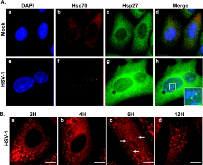
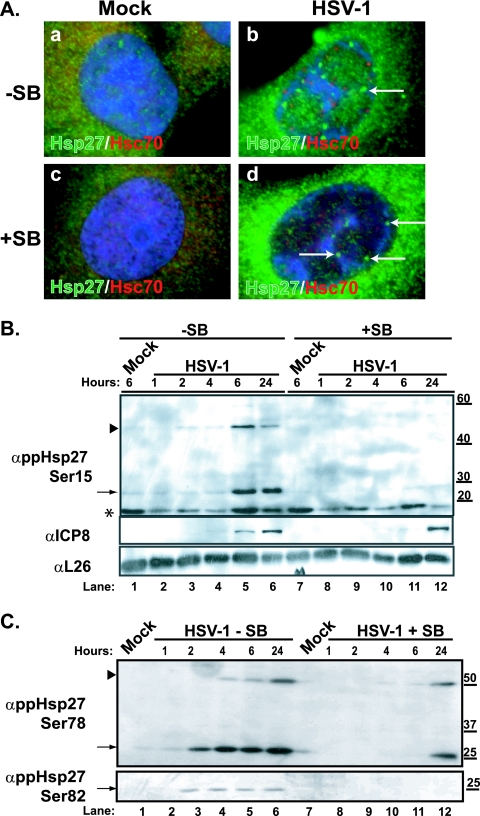
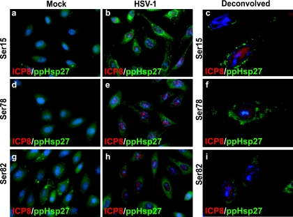
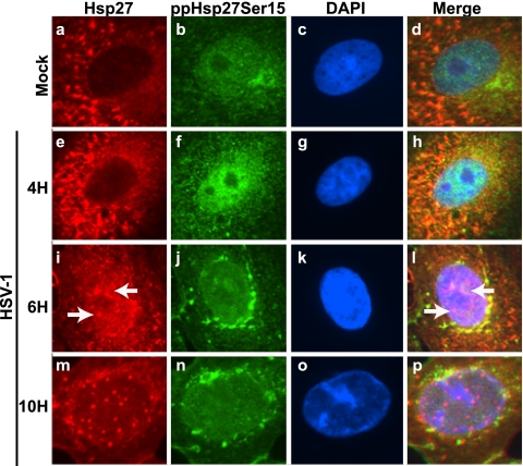
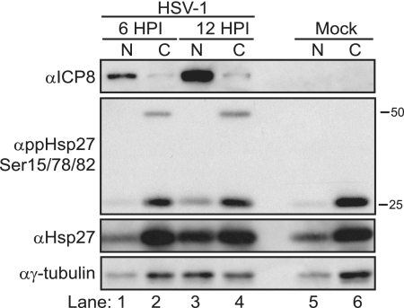
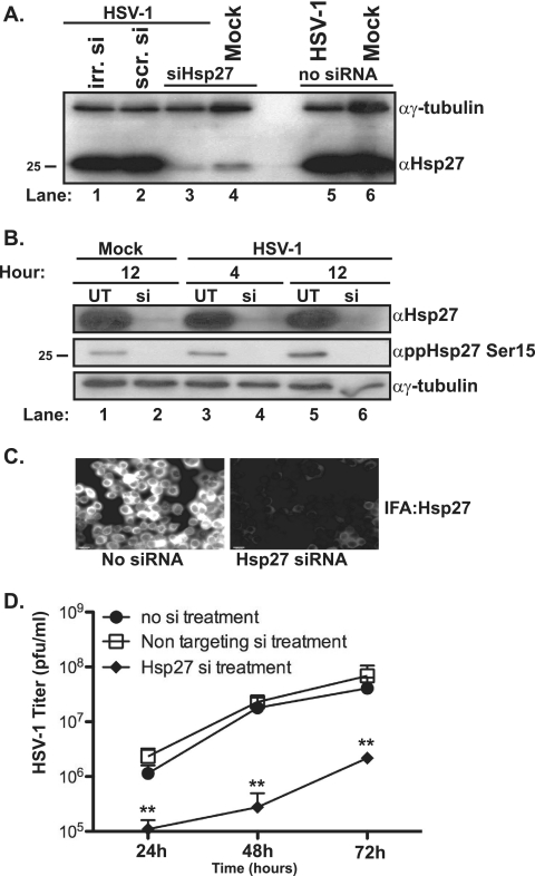
Chaperone-enriched domains are formed in the nuclei of cells lytically infected with herpes simplex virus type 1 (HSV-1). These domains, called VICE, for virus induced chaperone enriched, contain Hsc70, Hsp70, Hsp40, Hsp90, polyubiquitinated proteins, and components of the proteasome machinery. Accumulating evidence indicates that these sites may be utilized during infection to sequester misfolded, modified, or otherwise unwanted proteins away from viral replication compartments, sites of robust transcription, DNA synthesis, and capsid maturation. To further explore the role of cellular chaperones and VICE domains during HSV-1 infection, we have analyzed the cytoprotective chaperone Hsp27. Here we present evidence that Hsp27, which is known to possess several antioxidant functions, is rapidly reorganized and modified at early stages in response to HSV-1 infection and signaling from the mitogen-activated protein kinase p38. Immunofluorescence analysis and fractionation experiments reveal disparate subcellular localizations of nonphosphorylated and phosphorylated forms of Hsp27 during wild-type HSV-1 infection. Unmodified forms of Hsp27 are localized in nuclear foci that are outside of replication compartments, adjacent to VICE domains, and in the cytoplasm. Conversely, we find that phosphorylated forms of Hsp27 are localized exclusively in the cytoplasm. Last, in cells depleted of all forms of Hsp27, virus replication is significantly reduced.
Figures
Similar articles
-
Accumulation of oxidized proteins in Herpesvirus infected cells.Free Radic Biol Med. 2010 Aug 1;49(3):383-91. doi: 10.1016/j.freeradbiomed.2010.04.026. Epub 2010 May 2. Free Radic Biol Med. 2010. PMID: 20441790 Free PMC article.
-
The Herpes Simplex Virus 1 Immediate Early Protein ICP22 Is a Functional Mimic of a Cellular J Protein.J Virol. 2020 Jan 31;94(4):e01564-19. doi: 10.1128/JVI.01564-19. Print 2020 Jan 31. J Virol. 2020. PMID: 31748398 Free PMC article.
-
Virus-Induced Chaperone-Enriched (VICE) domains function as nuclear protein quality control centers during HSV-1 infection.PLoS Pathog. 2009 Oct;5(10):e1000619. doi: 10.1371/journal.ppat.1000619. Epub 2009 Oct 9. PLoS Pathog. 2009. PMID: 19816571 Free PMC article.
-
UBE1a Suppresses Herpes Simplex Virus-1 Replication.Viruses. 2020 Dec 4;12(12):1391. doi: 10.3390/v12121391. Viruses. 2020. PMID: 33291814 Free PMC article.
-
The Clinical Significance of Phosphorylated Heat Shock Protein 27 (HSPB1) in Pancreatic Cancer.Int J Mol Sci. 2016 Jan 21;17(1):137. doi: 10.3390/ijms17010137. Int J Mol Sci. 2016. PMID: 26805817 Free PMC article. Review.
Cited by
-
Accumulation of oxidized proteins in Herpesvirus infected cells.Free Radic Biol Med. 2010 Aug 1;49(3):383-91. doi: 10.1016/j.freeradbiomed.2010.04.026. Epub 2010 May 2. Free Radic Biol Med. 2010. PMID: 20441790 Free PMC article.
-
Bortezomib-induced unfolded protein response increases oncolytic HSV-1 replication resulting in synergistic antitumor effects.Clin Cancer Res. 2014 Jul 15;20(14):3787-98. doi: 10.1158/1078-0432.CCR-14-0553. Epub 2014 May 9. Clin Cancer Res. 2014. PMID: 24815720 Free PMC article.
-
AR-12 Inhibits Chaperone Proteins Preventing Virus Replication and the Accumulation of Toxic Misfolded Proteins.J Clin Cell Immunol. 2016 Oct;7(5):454. doi: 10.4172/2155-9899.1000454. Epub 2016 Sep 16. J Clin Cell Immunol. 2016. PMID: 27957385 Free PMC article. No abstract available.
-
Alteration in superoxide dismutase 1 causes oxidative stress and p38 MAPK activation following RVFV infection.PLoS One. 2011;6(5):e20354. doi: 10.1371/journal.pone.0020354. Epub 2011 May 31. PLoS One. 2011. PMID: 21655261 Free PMC article.
-
The kinase inhibitor SFV785 dislocates dengue virus envelope protein from the replication complex and blocks virus assembly.PLoS One. 2011;6(8):e23246. doi: 10.1371/journal.pone.0023246. Epub 2011 Aug 17. PLoS One. 2011. PMID: 21858043 Free PMC article.
References
-
- Arrigo, A. P. 2001. Hsp27: novel regulator of intracellular redox state. IUBMB Life 52:303-307. - PubMed
-
- Arrigo, A. P. 2007. The cellular “networking” of mammalian Hsp27 and its functions in the control of protein folding, redox state and apoptosis. Adv. Exp. Med. Biol. 594:14-26. - PubMed
-
- Arrigo, A. P., S. Virot, S. Chaufour, W. Firdaus, C. Kretz-Remy, and C. Diaz-Latoud. 2005. Hsp27 consolidates intracellular redox homeostasis by upholding glutathione in its reduced form and by decreasing iron intracellular levels. Antioxid. Redox Signal. 7:414-422. - PubMed
-
- Boros, S., B. Kamps, L. Wunderink, W. de Bruijn, W. W. de Jong, and W. C. Boelens. 2004. Transglutaminase catalyzes differential crosslinking of small heat shock proteins and amyloid-beta. FEBS Lett. 576:57-62. - PubMed
-
- Bruey, J. M., C. Ducasse, P. Bonniaud, L. Ravagnan, S. A. Susin, C. Diaz-Latoud, S. Gurbuxani, A. P. Arrigo, G. Kroemer, E. Solary, and C. Garrido. 2000. Hsp27 negatively regulates cell death by interacting with cytochrome c. Nat. Cell Biol. 2:645-652. - PubMed
Publication types
MeSH terms
Substances
Grants and funding
LinkOut - more resources
Full Text Sources
Medical
Research Materials
Miscellaneous
