. 2009 Sep;20(17):3878-87.
doi: 10.1091/mbc.e09-01-0038. Epub 2009 Jul 8.

Central role of the oxygen-dependent degradation domain of Drosophila HIFalpha/Sima in oxygen-dependent nuclear export

Affiliations

Central role of the oxygen-dependent degradation domain of Drosophila HIFalpha/Sima in oxygen-dependent nuclear export

Maximiliano Irisarri et al. Mol Biol Cell. 2009 Sep.

Abstract

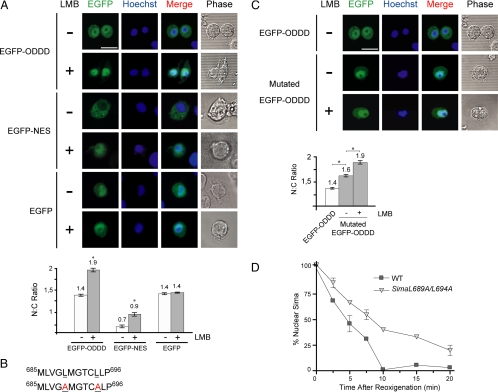
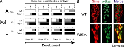
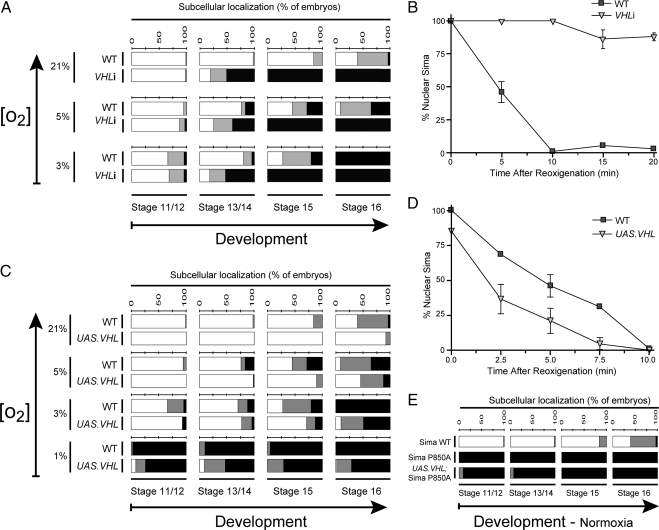
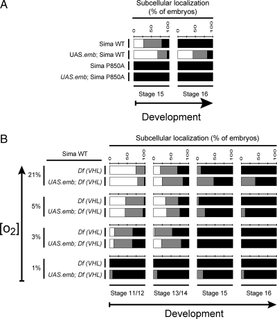
The Drosophila HIFalpha homologue, Sima, is localized mainly in the cytoplasm in normoxia and accumulates in the nucleus upon hypoxic exposure. We have characterized the mechanism governing Sima oxygen-dependent subcellular localization and found that Sima shuttles continuously between the nucleus and the cytoplasm. We have previously shown that nuclear import depends on an atypical bipartite nuclear localization signal mapping next to the C-terminus of the protein. We show here that nuclear export is mediated in part by a CRM1-dependent nuclear export signal localized in the oxygen-dependent degradation domain (ODDD). CRM1-dependent nuclear export requires both oxygen-dependent hydroxylation of a specific prolyl residue (Pro850) in the ODDD, and the activity of the von Hippel Lindau tumor suppressor factor. At high oxygen tension rapid nuclear export of Sima occurs, whereas in hypoxia, Sima nuclear export is largely inhibited. HIFalpha/Sima nucleo-cytoplasmic localization is the result of a dynamic equilibrium between nuclear import and nuclear export, and nuclear export is modulated by oxygen tension.


Figures

Figure 1.
Figure 1.
Sima nuclear export depends on the nuclear export receptor CRM1/Embargoed. (A) Categories of Sima subcellular localization in en-Gal4/UAS-Sima/UAS-nGFP.LacZ transgenic embryos: nuclear, ubiquitous, and cytoplasmic sample photographs of groups of cells overexpressing Sima in embryonic ectodermal cells (for quantitative criteria see Materials and Methods; see also Dekanty et al., 2005). (B) Sima becomes more nuclear in embargoed/CRM1 mutant embryos. □, cytoplasmic localization of Sima; ■, nuclear localization; and ▩, ubiquitous localization. Sima subcellular localization in wild-type embryos (WT) was progressively more nuclear as embryogenesis proceeds and gradually more nuclear as oxygen levels decrease (χ2 test; p < 10−4; n > 50). Nuclear localization increased in embargoed (emb3) homozygous mutant embryos (p < 10−3; n > 50) at 21, 5, and 3% O2 concentrations. At 1% O2 Sima was already totally nuclear in wild-type embryos throughout development. (C) Nuclear export assay of Sima protein upon reoxygenation. Stage 16 embryos expressing Sima protein were exposed to 1% O2 during 4 h and then transferred to normoxia and stained for Sima at different time points after reoxygenation. In WT embryos Sima is totally exported from the nucleus in only 10 min, whereas in emb3 mutants, Sima is exported at a slower rate (Kaplan-Meier; p < 10−4; n > 30). (D) Cytoplasmic localization of Sima was enhanced in embryos overexpressing the Emb protein (p < 10−4; n > 40) at all tested oxygen concentrations throughout embryogenesis, suggesting that increased levels of CRM1 promote nuclear export of Sima protein. No differences occurred at embryonic stages 11–12 in normoxia or 5% O2, as in wild-type embryos (y w), Sima was already fully cytoplasmic in these conditions. (E) Sima nuclear export is faster in embryos overexpressing CRM1/Embargored. Nuclear export assays were carried out as in C, revealing that upon expression of the Embargoed protein in transgenic embryos, Sima is exported more efficiently (Kaplan-Meier; p < 10−4; n > 30). (F) Sima transcriptional activity depends on the rate of nuclear export. A Sima-responsive LDH-LacZ Sima-responsive transcriptional reporter (Lavista-Llanos et al., 2002) was crossed into the UAS lines expressing Sima in embryos that were otherwise wild-type (WT), mutant for CRM1/embargoed (emb3), or overexpressing the Emb protein (UAS.emb), and β-galactosidase activity was determined through a colorimetric assay (Materials and Methods). Activity of the Sima-dependent LacZ reporter was significantly increased in emb3 mutant embryos and reduced in embryos overexpressing the Emb protein (Student's t test; * p < 10−3; n = 3).
Figure 2.
Figure 2.
Identification of a functional nuclear export signal (NES) in the Sima oxygen-dependent degradation domain (ODDD). (A) Sensitivity of an EGFP-ODDD construct to leptomycin B (LMB). An EGFP-ODDD (amino acids 665–871) fusion protein, an EGFP-NES–positive control or EGFP alone were transfected to S2 cells, and subcellular localization of the constructs was analyzed by confocal microscopy 2 h after addition or not of 30 mM LMB; the nuclear:cytoplasmic (N–C) fluorescence ratio was calculated and is shown in the bottom panel. Although EGFP-ODDD and the EGFP-NES–positive control became clearly more nuclear upon addition of LMB (Student's t test; significant difference: * p < 10−7; n > 20), EGFP was insensitive to LMB, suggesting that a functional CRM1-dependent NES occurs in Sima ODDD. (B) Amino acid sequence of the NES of the Sima ODDD. Top line, wild-type sequence; bottom line mutagenized sequence in which two leucine residues were replaced by alanines (underlined). (C) The NES of the ODDD promotes nuclear export of an EGFP reporter. EGFP fusions were generated with the wild-type or the mutagenized version of the NES. The chimera including the mutagenized NES was remarkably more nuclear than the one with the wild-type NES; treatment with LMB rendered the construct even more nuclear (Student's t test; significant difference: * p < 10−7; n > 20), suggesting that the mutations provoked reduction but not complete elimination of NES function. The N–C fluorescence ratios of these experiments are shown in the bottom panel. (D) A full-length Sima protein, mutagenized at the NES of the ODDD as in B, is exported at a slower rate than wild-type Sima in transgenic embryos, in a reoxygenation assay similar to the one described in Figure 1.
Figure 3.
Figure 3.
Prolyl hydroxylation is essential for Sima nuclear export. (A) In fatiga1 homozygous mutant embryos, Sima was more nuclear than in the wild-type controls (WT; p < 10−4; n > 50) at all tested oxygen levels throughout embryogenesis, indicating that prolyl hydroxylation is necessary for Sima cytoplasmic localization. The experimental design is the same as in Figure 1, B and D. ■, nuclear; ▩, ubiquitous; and □, cytoplasmic. (B) Wild-type Sima protein (red) was predominantly cytoplasmic in normoxia (cell nuclei are marked by anti-βgal staining; green); when Sima proline 850 was replaced by an alanine (P850A), Sima became constitutively nuclear with no signal detected in the cytoplasm. Scale bar, 10 μm.
Figure 4.
Figure 4.
VHL promotes cytoplasmic localization of Sima. (A) In embryos expressing a VHL RNAi transgene (VHLi), Sima was more nuclear than in wild-type (WT) controls at all tested oxygen levels after stage 13 (p < 10−4; n > 40). ■, nuclear; ▩, ubiquitous; and □, cytoplasmic. (B) Sima nuclear export assay upon reoxygenation, in embryos expressing VHLi, reveals that Sima export was severely impaired (Kaplan-Meier; p < 10−4; n > 30). (C) Overexpression of Drosophila VHL increased Sima cytoplasmic localization at all tested oxygen levels throughout embryogenesis with the exception of those conditions where in the wild-type controls, Sima was already fully cytoplasmic (p < 10−2; n > 40). (D) Nuclear export of Sima is more efficient in embryos overexpressing VHL. Sima nuclear export assay similar to the one depicted in B (Kaplan-Meier; p < 10−4; n > 30). (E) A mutagenized version of Sima in which proline 850 was replaced by an alanine (SimaP850A) was insensitive to VHL overexpression, remaining totally nuclear even in normoxia (n > 40).
Figure 5.
Figure 5.
Prolyl hydroxylation and VHL function are required for CRM1-dependent nuclear export of Sima. (A) Overexpression of the nuclear export receptor CRM1/Embargoed enhanced the rate of Sima nuclear export, leading to increased levels of wild-type Sima protein in the cytoplasm in normoxia (p < 10−4; n > 40) but failed to enhance nuclear export of SimaP850A, which remained localized exclusively in the nuclear compartment even in normoxia (n > 40). (B) Sima subcellular localization in VHL loss-of-function embryos [Df (VHL) homozygous for the Df(2R)en-A deficiency] remained largely unchanged upon overexpression of CRM1/Embargoed, revealing that VHL function is required for Sima nuclear export (n > 50). ■, nuclear; ▩, ubiquitous; and □, cytoplasmic.
Figure 6.
Figure 6.
Model for oxygen-dependent regulation of Sima subcellular localization and degradation. Sima is constitutively imported into the nucleus. In hypoxia Sima forms a heterodimer with the β-subunit Tango, and induces transcription; in normoxia the Prolyl 850 residue is hydroxylated by the PHD Drosophila homologue Fatiga, ubiquitinated by VHL and exported to the cytoplasm in a CRM1-dependent manner. Prolyl-hydroxylation, ubiquitination, and degradation at the 26S proteasome can presumably occur both in the nucleus and the cytoplasm (Berra et al., 2001).

Similar articles

Cited by

References

    1. Adryan B., Decker H. J., Papas T. S., Hsu T. Tracheal development and the von Hippel-Lindau tumor suppressor homolog in Drosophila. Oncogene. 2000;19:2803–2811. - PubMed
    1. Arquier N., Vigne P., Duplan E., Hsu T., Therond P. P., Frelin C., D'Angelo G. Analysis of the hypoxia-sensing pathway in Drosophila melanogaster. Biochem. J. 2006;393:471–480. - PMC - PubMed
    1. Bacon N. C., Wappner P., O'Rourke J. F., Bartlett S. M., Shilo B., Pugh C. W., Ratcliffe P. J. Regulation of the Drosophila bHLH-PAS protein Sima by hypoxia: functional evidence for homology with mammalian HIF-1 alpha. Biochem. Biophys. Res. Commun. 1998;249:811–816. - PubMed
    1. Berra E., Roux D., Richard D. E., Pouyssegur J. Hypoxia-inducible factor-1 alpha (HIF-1 alpha) escapes O(2)-driven proteasomal degradation irrespective of its subcellular localization: nucleus or cytoplasm. EMBO Rep. 2001;2:615–620. - PMC - PubMed
    1. Bogerd H. P., Fridell R. A., Benson R. E., Hua J., Cullen B. R. Protein sequence requirements for function of the human T-cell leukemia virus type 1 Rex nuclear export signal delineated by a novel in vivo randomization-selection assay. Mol. Cell. Biol. 1996;16:4207–4214. - PMC - PubMed

Publication types

MeSH terms

Substances

LinkOut - more resources