Review
. 2009 Dec;297(6):F1477-501.
doi: 10.1152/ajprenal.00327.2009. Epub 2009 Jul 8.

Cell biology and physiology of the uroepithelium

Affiliations
Review

Cell biology and physiology of the uroepithelium

Puneet Khandelwal et al. Am J Physiol Renal Physiol. 2009 Dec.

Abstract

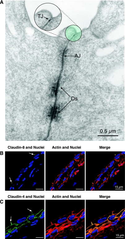
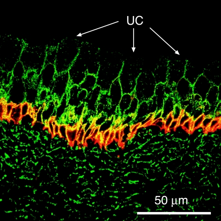
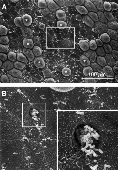
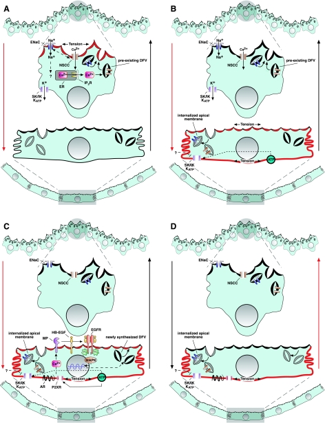
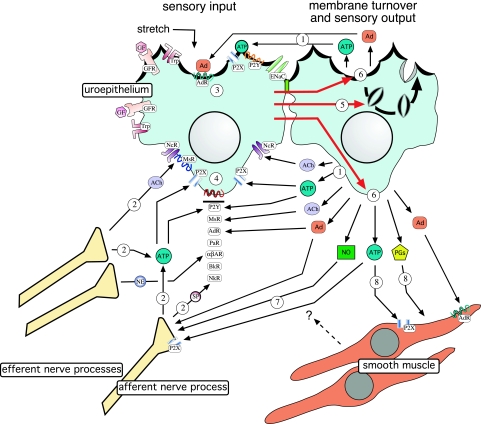
The uroepithelium sits at the interface between the urinary space and underlying tissues, where it forms a high-resistance barrier to ion, solute, and water flux, as well as pathogens. However, the uroepithelium is not simply a passive barrier; it can modulate the composition of the urine, and it functions as an integral part of a sensory web in which it receives, amplifies, and transmits information about its external milieu to the underlying nervous and muscular systems. This review examines our understanding of uroepithelial regeneration and how specializations of the outermost umbrella cell layer, including tight junctions, surface uroplakins, and dynamic apical membrane exocytosis/endocytosis, contribute to barrier function and how they are co-opted by uropathogenic bacteria to infect the uroepithelium. Furthermore, we discuss the presence and possible functions of aquaporins, urea transporters, and multiple ion channels in the uroepithelium. Finally, we describe potential mechanisms by which the uroepithelium can transmit information about the urinary space to the other tissues in the bladder proper.


Figures

Fig. 1.
Fig. 1.
Tissue architecture of the uroepithelium. Rat bladder uroepithelium was stained with FITC-phalloidin (green) to label the cortical actin cytoskeleton associated with plasma membranes of uroepithelial cells, TO-PRO3 (blue) to label nuclei, and an antibody that recognizes the cytoplasmic domain of uroplakin (UP) IIIa (red). Left: distribution of UPIIIa and nuclei. Middle: actin and nuclei. Umbrella cells are marked by arrows, intermediate cells by yellow circles, and basal cells by white circles. A capillary (c) below the basal cell layer is shown. Right: all 3 markers.
Fig. 2.
Fig. 2.
Ultrastructure of bladder uroepithelium. A: scanning electron micrographs of mucosal surface of the bladder. Left: overview of the apical surface of rabbit umbrella cells. Borders of an individual umbrella cell are indicated by arrowheads. Right: ridge, consisting of zippered apical membrane (ZM), at the periphery of adjacent umbrella cells. B: transmission electron micrograph of the apical pole of a rat umbrella cell. Examples of discoidal/fusiform-shaped vesicles (DFV) are marked with arrows and hinges with arrowheads. Plaques are located in the intervening membrane between hinges. Apical cytoplasm of rat umbrella cells has disc-shaped (*) and fusiform-shaped vesicles. Multivesicular bodies (MVB; i.e., late endosomes) are indicated by arrows. C: asymmetric unit membrane (AUM) at the apical surface of a rat umbrella cell. Contrast was adjusted using Photoshop.
Fig. 3.
Fig. 3.
Structure of AUM particles and UP subunits. A: 3-dimensional reconstruction of an AUM particle as determined by electron cryomicroscopy. Inner ring consists of UPIa/UPII heterodimers (arrowhead), and outer ring consists of UPIb/UPIIIa heterodimers (arrow). Scale bar, 2 nm. [From Min et al. (146).] B: schematic depiction of interactions between UPIa/UPII and UPIb/UPIIIa heterodimers. Extracellular loop (EC)-2 of UPIa or UPIb interacts with the extracellular domain of UPII or UPIIIa, respectively. Green circles show sites of N-linked glycosylation. Transmembrane domains of UPII and UPIIIa/IIIb contain a conserved region (red) that extends to the extracellular domain of UPIIIa/UPIIIb. Sequence of the extracellular region of the conserved domain of UPIIIa is shown in single amino acid code. Region of the extracellular domain of UPIIIb (yellow) is >90% identical to a portion of human DNA mismatch repair enzyme-related PMSR6 protein. [From Birder et al. (24).] C: interactions between heterodimers in inner and outer rings of the AUM particle (left) and between adjacent UPIa/UPII heterodimers (right). [From Birder et al. (24).]
Fig. 4.
Fig. 4.
Trajectorial cytokeratin network of umbrella cells. A: 3-dimensional reconstruction of cytokeratin-20 network (red) at the apical pole of umbrella cells. Borders of adjacent umbrella cells are marked by the tight junction protein ZO-1 (green), and nuclei are shown in blue. [From Veranic et al. (212). Reprinted by permission of Springer Science+Business Media.] B: UP-positive DFV (green) are localized within tunnels formed by the cytokeratin-20-positive network (red). [From Veranic and Jezernik (216). Reprinted by permission of John Wiley & Sons, Inc.]
Fig. 5.
Fig. 5.
Claudin expression in rat bladder uroepithelium. A: junctional complex of the umbrella cell. Positions of tight junction (TJ), adherens junction (AJ), and desmosomes (Ds) are indicated with arrows. Inset: higher-magnification view of the tight junction. Contrast was adjusted with Photoshop. B: claudin-8 (green) is expressed at the apicolateral junction of the umbrella cell layer. Phalloidin staining (red) demarcates cell borders of the uroepithelium. C: claudin-4 (green) is associated with the basolateral surface of umbrella cells and plasma membranes of intermediate and basal cell layers. Arrows in B and C indicate location of the tight junction in the umbrella cell layer.
Fig. 6.
Fig. 6.
Aquaporin (AQP)-3 and urea transporter (UT)-B distribution in uroepithelium. UT-B (green) is distributed on the basolateral surface of dog bladder umbrella cells and is present on plasma membranes of intermediate and basal cell layers. AQP-3 (red) shares a similar distribution but is heavily expressed in the basal cell layer. Arrows, apical surface of representative umbrella cells (UC). [From Spector et al. (192).]
Fig. 7.
Fig. 7.
Association of uropathogenic Escherichia coli with bladder uroepithelium. A: E. coli adherent to the surface of the uroepithelium. Apical surfaces of umbrella cells flatten in response to bacterial adhesion, whereas adjacent umbrella cells with few adherent bacteria (*) maintain a dome-shaped morphology. B: higher-magnification view of bacterial adherence to umbrella cells and invasion between cells shown in boxed region in A. Higher-magnification view of boxed region in B shows site of bacterial invasion (inset).
Fig. 8.
Fig. 8.
Model for exocytic and endocytic responses to bladder filling and voiding. A: as the bladder fills, the mucosal surface unfurls and increased tension at the apical pole of the umbrella cell triggers opening of a nonselective cation channel (NSCC), stimulating influx of extracellular Ca2+. Increased intracellular Ca2+ concentration triggers Ca2+-dependent Ca2+ release from inositol trisphosphate (IP3)-dependent stores, which triggers the early-stage exocytic response. This response is likely driven by fusion of a preexisting pool of DFV with the apical plasma membrane of the umbrella cell. Exocytosis may amplify the initial response by stimulating delivery of additional stretch-sensing channels to the apical surface of the cell. Epithelial Na+ channel (ENaC), perhaps acting upstream of the NSCC, is also opened and may regulate the exocytic response by changing the membrane potential or the driving force for the entry of other ions. IP3R, IP3 receptor; SK/IK, small/intermediate-conductance K+ channels; KATP, ATP-sensitive K+ channel. B: as the epithelium bows further, outward tension in the basolateral membrane increases, stimulating stretch-induced endocytosis. Enhanced endocytosis would modulate the exocytic response by stimulating internalization of apical membrane channels and other sensory molecules (not shown). The mechanosensor at the basolateral membrane is unknown but may include integrins, the cytoskeleton, or stretch-modulated K+ channel(s), which may close in response to the increased tension or other intracellular mediators such as ATP. C: as the bladder fills and tension is present at mucosal and serosal surfaces of the umbrella cell, heparin-binding epidermal growth factor (HB-EGF) is cleaved at the apical surface of the umbrella cell by an unknown metalloproteinase (MP) and then binds to and activates apical EGF receptors (EGFR). In turn, EGFR activates ERK and p38 MAPK, which trigger the late-stage exocytic response by promoting new protein synthesis and, possibly, biosynthesis of new DFV. Transactivation of the EGFR may occur in response to ATP that is released upon stretch and binds to purinergic receptors (P2XR), stimulating an increase in the intracellular Ca2+ concentration. Adenosine, acting through adenosine receptors (AR), may also act by stimulating transactivation of the EGFR. D: during voiding, tension would be released from the apical surface of umbrella cells but would increase in the basolateral membrane as the detrusor contracts and actively pushes the mucosa toward the lumen of the bladder. Increased tension would further stimulate endocytosis of the apical membrane and its constituents, readying the umbrella cell for the next cycle of filling. Red arrows at periphery indicate bladder filling (A–C) or voiding (D). For clarity, we do not include underlying cell layers or connective tissue; however, umbrella cells likely transmit tension to the interconnected matrix, intermediate cells, and basal cells, and they, in turn, may impact or promote events in the overlying umbrella cell layer. [Adapted from Yu et al. (234).]
Fig. 9.
Fig. 9.
Bidirectional communication between the uroepithelium and other tissues in the uroepithelium-associated sensory web. The uroepithelium receives “sensory input” in a number of different forms, including mechanical stretch as the bladder fills and the release of soluble mediators such as ACh, adenosine, and ATP from the uroepithelium (step 1), adjacent afferent/efferent nerve processes (step 2), and, possibly, the smooth muscle or myofibroblasts (not shown). In turn, soluble mediators bind to cell surface receptors on the apical surface of umbrella cells (step 3), basolateral surfaces of umbrella cells (step 4), and plasma membranes of underlying intermediate and basal cells (not shown). Receptor binding or channel activation results in changes in the uroepithelium, including membrane turnover at the apical plasma membrane of the umbrella cell (and, possibly, at other plasma membrane domains of the uroepithelium; step 5) and release of mediators such as ACh, adenosine, ATP, nitric oxide (NO), and prostaglandins (PGs; step 6), which act as “sensory outputs.” These outputs can act in an autocrine manner to further alter uroepithelial function (step 1), or they can bind to receptors on sensory afferent nerve processes (step 7) and/or the detrusor muscle (step 8) to regulate nerve and muscle function. α,β-AR, α,β-adrenergic receptor; Ad, adenosine; AdR, adenosine receptor; Bk, bradykinin; BkR, bradykinin receptor; GF, growth factor; GFR, growth factor receptor; MsR, muscarinic receptor; NcR, nicotinic receptor; NkR, neurokinin receptor; PaR, proteinase-activated receptor; Pr, proteinase; Trp, transient receptor potential channel family member. [Adapted from Apodaca et al. (11).]

References

    1. Aboushwareb T, Zhou G, Deng FM, Turner C, Andersson KE, Tar M, Zhao W, Melman A, D'Agostino R, Jr, Sun TT, Christ GJ. Alterations in bladder function associated with urothelial defects in uroplakin II and IIIa knockout mice. Neurourol Urodyn In press - PMC - PubMed
    1. Acharya P, Beckel J, Wang E, Ruiz W, Rojas R, Apodaca G. Distribution of the tight junction proteins ZO-1, occludin, and claudin-4, -8, and -12 in bladder epithelium. Am J Physiol Renal Physiol 287: F305–F318, 2004 - PubMed
    1. Adachi W, Okubo K, Kinoshita S. Human uroplakin Ib in ocular surface epithelium. Invest Ophthalmol Vis Sci 41: 2900–2905, 2000 - PubMed
    1. Alenghat FJ, Nauli SM, Kolb R, Zhou J, Ingber DE. Global cytoskeletal control of mechanotransduction in kidney epithelial cells. Exp Cell Res 301: 23–30, 2004 - PubMed
    1. Alroy J, Merk FB, Morre DJ, Weinstein RS. Membrane differentiation in the Golgi apparatus of mammalian urinary bladder epithelium. Anat Rec 203: 429–440, 1982 - PubMed

Publication types

LinkOut - more resources