. 2009 Jun;88(6):539-44.
doi: 10.1177/0022034509338008.

Function of beta1 integrin in oral epithelia and tooth bud morphogenesis

Affiliations

Function of beta1 integrin in oral epithelia and tooth bud morphogenesis

B Chen et al. J Dent Res. 2009 Jun.

Abstract

Integrin beta1 is critical for basement membrane organization and hair follicle morphogenesis in the skin epidermis; however, less is known about its function in the developing oral epithelium. Since the skin and oral epithelia share structural similarity, we hypothesized that beta1 integrin function would be critical for the normal development of oral epithelium and tooth buds. The conditional (oral mucosa-specific) beta1 integrin knockout (KO) mice displayed severe disruption of the basement membrane of the tongue epithelium and developing tooth buds. Interestingly, unlike the developing hair follicles, early morphological development of the KO molar tooth buds was normal. However, subsequent morphogenetic events, such as cusp formation, cervical loop down-growth, and ameloblast polarization, did not occur normally. Primary KO oral keratinocytes showed defective cell spreading and robust focal adhesions. Our studies indicate that beta1 integrin plays an essential role in the normal development of the oral epithelium and its appendages.


Figures

Figure 1.
Figure 1.
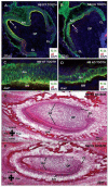
Consequences of the loss of β1 integrin in the oral mucosa. Sagittal sections of the tongues of newborn mice were stained with H&E. (A) The tongue epithelium of the WT mouse was intact, and (B) In the β1 KO, the epithelia separated from the underlying mesenchyme (arrowheads). Coronal sections of embryonic day (E) 17.5 WT and KO embryos were stained with antibodies to β1 integrin (green) and laminin 5 (red) (C,D), K-14 (green) and tenascin C (red) (E,F), and β6 integrin (green) and laminin 5 (red) (G,H). Nuclei were stained with DAPI (blue) in all sections. (C) β1 integrin was expressed in both the epithelia and mesenchyme of the tongue. Basement membrane of the WT tongue and palate was intact. (D) In contrast, in the β1 integrin KO, the expression of β1 integrin was lost only from the tongue epithelia, but was still expressed in the mesenchyme. The basement membrane of the tongue, as judged by the expression of laminin 5, was disrupted. Tenascin C was up-regulated in the KO (F), and there is a de novo expression of β6 integrin in the KO (G). BM, basement membrane; P, palate; T, tongue; TE, tongue epithelia; TM, tongue mesenchyme.
Figure 2.
Figure 2.
Development of ameloblasts in molar and incisor tooth buds. Coronal sections of NB WT and β1 KO tooth buds were labeled with K-14 (green), α6 integrin (red), and DAPI (blue). (A) The WT tooth bud was in the late bell stage, and distinct cusps and cervical loops could be seen. (B) The KO tooth bud was in the mid-bell stage. The cusps were not as distinct as in WT, and the cervical loops did not grow down as far. (C) In the WT tooth bud, the developing ameloblasts were elongated and well-polarized. (D) In the KO tooth bud, the developing ameloblasts were rounded and not polarized. (E-F) Sagittal sections of developing newborn lower incisors were processed by H&E. (E) In the developing WT lower incisor, the ameloblasts were elongated and polarized, and the cervical loops (where the epithelial stem cells are located) were distinct. (F) In the developing KO lower incisor, the ameloblasts were not elongated or polarized, and the cervical loops were not well-defined (asterisk in F). Am, ameloblasts; C, cusp; DP, dental papilla; BM, basement membrane; CL, cervical loops; Od, odontoblasts.
Figure 3.
Figure 3.
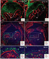
Basement membrane defects and loss of proliferation in the KO tooth buds. Coronal sections of E17.5 and NB, WT, and KO embryos were stained with antibodies to e-cadherin (green) and collagen IV (red) (A,B) and the proliferation marker phosphohistone H3 [PH3] (green) and HSPG (red) (C,D). (A) The basement membrane of the WT tooth bud was intact, whereas (B) the basement membrane surrounding the developing β1 KO tooth bud was severely disrupted, particularly around the cervical loops (arrowheads in B). (C) Most of the developing ameloblasts were labeled with PH3 in the WT, and the expression of HSPG was distinct around the developing cusps. (D) The number of proliferating ameloblasts, as judged by PH3 staining, was reduced in the KO, and the expression of HSPG, especially around the cusps, was not distinct. (C’) Higher-magnification image of the boxed area in C. Several PH3-positive cells could be seen, and there was strong expression of HSPG. (D’) Higher-magnification image of the boxed area in D. Very few proliferating ameloblasts could be seen, and the expression of HSPG was weak. OE, outer dental epithelia; IE, inner dental epithelia; BM, basement membrane; CL, cervical loop; Am, ameloblasts; DP, dental papilla.
Figure 4.
Figure 4.
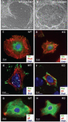
Isolation of WT and KO keratinocytes: (A) β1 WT and (B) KO cells were co-cultured with fibroblast feeders in media that contained 1.2 mM calcium. The WT and KO cells were plated on fibronectin-coated coverslips and labeled with the following antibodies: vinculin, paxillin, and K5 (red in E,F and green in G,H), TRITC-phalloidin, and α6 integrin (red), and the nuclei were counterstained with DAPI. The KO cells (D,F,H) were half as well-spread as WT keratinocytes (C,E,G), and had robust focal adhesions compared with WT cells. The focal complexes that could be seen in the center of the WT cells (arrowheads in E) were not formed in the KO. The keratin cytoskeleton was not affected (H).

References

    1. Ahmed N, Riley C, Rice GE, Quinn MA, Baker MS. (2002). Alpha(v)beta(6) integrin-A marker for the malignant potential of epithelial ovarian cancer. J Histochem Cytochem 50:1371-1380 - PubMed
    1. Arihiro K, Kaneko M, Fujii S, Inai K, Yokosaki Y. (2000). Significance of alpha 9 beta 1 and alpha v beta 6 integrin expression in breast carcinoma. Breast Cancer 7:19-26 - PubMed
    1. Berrier AL, Yamada KM. (2007). Cell-matrix adhesion. J Cell Physiol 213:565-573 - PubMed
    1. Brakebusch C, Grose R, Quondamatteo F, Ramirez A, Jorcano JL, Pirro A, et al. (2000). Skin and hair follicle integrity is crucially dependent on beta 1 integrin expression on keratinocytes. EMBO J 19:3990-4003 - PMC - PubMed
    1. Breuss JM, Gallo J, DeLisser HM, Klimanskaya IV, Folkesson HG, Pittet JF, et al. (1995). Expression of the beta 6 integrin subunit in development, neoplasia and tissue repair suggests a role in epithelial remodeling.J Cell Sci 108(Pt 6):2241-2251 - PubMed

Publication types