. 2009 Jul 21;106(29):11995-2000.
doi: 10.1073/pnas.0905188106. Epub 2009 Jul 8.

Polysialic acid governs T-cell development by regulating progenitor access to the thymus

Affiliations

Polysialic acid governs T-cell development by regulating progenitor access to the thymus

Penelope M Drake et al. Proc Natl Acad Sci U S A. .

Abstract

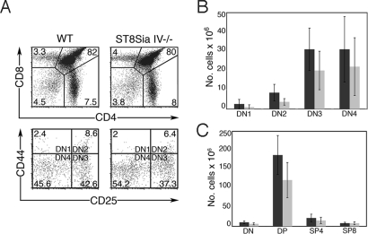
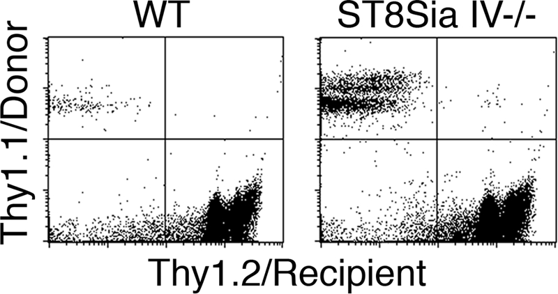
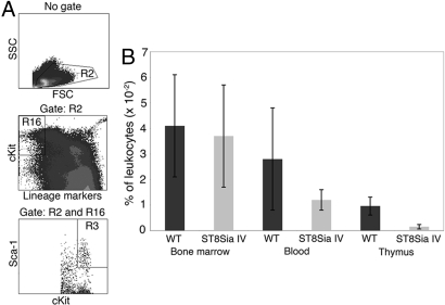
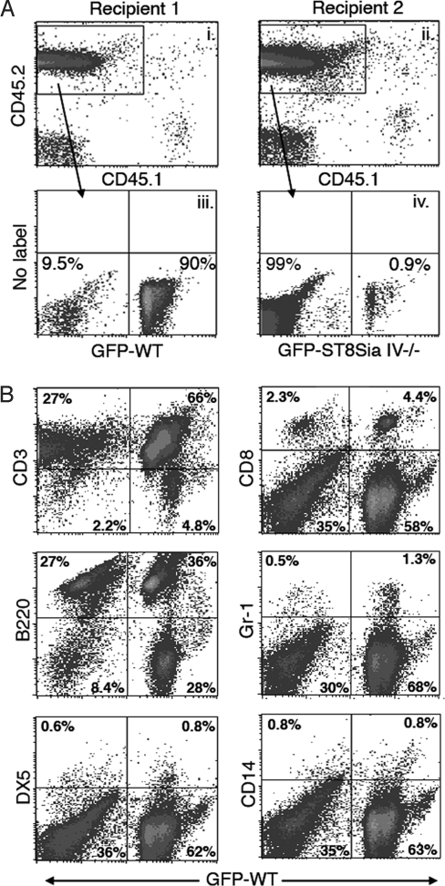
Although the polysialyltransferase ST8Sia IV is expressed in both primary and secondary human lymphoid organs, its product, polysialic acid (polySia), has been largely overlooked by immunologists. In contrast, polySia expression and function in the nervous system has been well characterized. In this context, polySia modulates cellular adhesion, migration, cytokine response, and contact-dependent differentiation. Provocatively, these same processes are vital components of immune development and function. We previously established that mouse multipotent hematopoietic progenitors use ST8Sia IV to express polySia on their cell surfaces. Here, we demonstrate that, relative to wild-type controls, ST8Sia IV(-/-) mice have a 30% reduction in total thymocytes and a concomitant deficiency in the earliest thymocyte precursors. T-cell progenitors originate in the bone marrow and are mobilized to the blood at regular intervals by unknown signals. We performed in vivo reconstitution experiments in which ST8Sia IV(-/-) progenitors competed with wild-type cells to repopulate depleted or deficient immune subsets. Progenitors lacking polySi exhibited a specific defect in T-cell development because of an inability to access the thymus. This phenotype probably reflects a decreased capacity of the ST8Sia IV(-/-) progenitors to escape from the bone marrow niche. Collectively, these results provide evidence that polySia is involved in hematopoietic development.


Conflict of interest statement

The authors declare no conflict of interest.

Figures

Fig. 1.
Fig. 1.
ST8Sia IV−/− mice have decreased numbers of DN and DP subsets. Wild-type (WT) and ST8Sia IV−/− thymocytes were analyzed by flow cytometry. (A) Representative flow cytometry plots of thymocyte populations from individual WT and ST8Sia IV−/− mice. CD4 and CD8 expression defines the DN, DP, SP4, and SP8 populations (Top), whereas CD44 and CD25 expression identifies the DN1–4 subsets (Bottom). The numbers in the panels indicate the percentages comprising each population for the experiment shown. Refer to Table 1 for the mean percentage and SD of each subset. (B) Total cell numbers in DN, DP, SP4, and SP8 and (C) DN1–DN4 thymic populations were assessed in 18 WT (dark gray bars) and 16 ST8Sia IV−/−mice (light gray bars). Statistically significant decreases were noted in ST8Sia IV−/− animals in the DN1 (P = 0.04), DN2 (P = 0.0004), DN3 (P = 0.01), total DN (P = 0.003), and DP (P = 0.0004) subsets.
Fig. 2.
Fig. 2.
ST8Sia IV−/− mice have empty progenitor niches. Thy1.2+ WT and ST8Sia IV−/− recipients were injected with exogenous donor bone marrow cells, and 3 weeks later thymocytes were analyzed for transferred cells. As expected, WT recipients showed minimal engraftment (Left; 0.47% ± 0.48%, n = 6). In contrast, ST8Sia IV−/− thymocytes consistently contained a significant contribution from donor cells (Right; 11.4% ± 5.4%, n = 7; P = 0.0005). A representative example from 1 WT and 1 ST8Sia IV−/− recipient is shown.
Fig. 3.
Fig. 3.
The percentage of early T-lineage progenitors is decreased in the ST8Sia IV−/− thymus. (A) Bone marrow, blood, and thymocytes from age-matched WT and ST8Sia IV−/− (ST8Sia IV) mice were analyzed by flow cytometry for LSKs in the bone marrow, blood, and thymus.(B) The prevalence of LSKs in each organ was assessed as the percentage of total leukocytes. A 6-fold decrease in the percentage of LSKs was observed in ST8Sia−/− thymocytes relative to controls (P < 0.000003).
Fig. 4.
Fig. 4.
ST8Sia IV−/− mice have a T-cell developmental defect. Irradiated CD45.1+ recipient mice were injected with equal numbers of congenic CD45.2+ wild-type and CD45.2+ ST8Sia IV−/− bone marrow cells. In each experiment, 1 of the donor animals (either wild type or ST8Sia IV−/−) also expressed GFP, as a marker of cell origin. Four months after injection, thymocytes (A) and splenocytes (B) were analyzed by flow cytometry. (A) Data from 2 representative recipient mice that received bone marrow transfers containing either wild-type:GFP (Recipient 1) or ST8SiaIV−/−:GFP (Recipient 2) are shown. (i and ii) Thymocyte CD45.1/CD45.2 expression was determined, and donor-derived (CD45.2+) cells were gated for further analyses. (iii) GFP+ cells, derived from wild-type precursors in this experiment, comprised 90% of all CD45.2+ thymocytes, whereas ST8Sia IV−/− descendants (CD45.2+/GFP) accounted for 9.5%. (iv) GFP+ cells, derived from ST8Sia IV−/− precursors in this experiment, comprised less than 1% of all CD45.2 thymocytes, whereas wild-type descendants (CD45.2+/GFP) accounted for 99%. (B) The representative splenocyte subset data shown were obtained from Recipient 1 (Panel A), which received donor bone marrow containing wild-type GFP+ progenitors. Splenocytes were analyzed for CD45.2/CD45.1 expression (data not shown), and CD45.2+ cells were gated for further analyses. A panel of antibodies against hematopoietic lineage markers was used to determine the contribution of wild-type and ST8Sia IV−/− donors to various leukocyte populations; co-expression of these antigens and GFP was assessed. Numbers represent the percentage of cells in each quadrant. Note that whereas the thymus of Recipient 1 was largely repopulated by GFP+ wild-type-derived cells (Panel A), the spleen of this animal contained significant numbers of GFP ST8Sia IV−/−-derived cells (compare right and left quadrants of flow plots). Overall, combined data from all recipient mice revealed that both donors made similar contributions to the B-cell (B220), NK-cell (DX5), and monocyte (CD14) populations; however, wild-type progenitors yielded about twice as many neutrophils (Gr-1) and about 5 times as many T cells (CD8) as did ST8Sia IV−/− precursors (data not shown).
Fig. 5.
Fig. 5.
ST8Sia IV−/− progenitors do not traffic effectively to the thymus. Bone marrow from Thy1.1 congenic WT (dark gray bars) and GFP-expressing ST8Sia IV−/− mice (light gray bars) was injected 1:1 either intravenously (IV) or intrathymically (IT) into congenic Thy1.2 IL-7Rα−/− recipient mice. Three weeks after injection, thymocytes were analyzed by flow cytometry to determine donor origin. When injected intravenously, ST8Sia IV−/− progenitors did not repopulate the thymus as efficiently as did WT cells (*, P < 0.02).

References

    1. Rossi FM, et al. Recruitment of adult thymic progenitors is regulated by P-selectin and its ligand PSGL-1. Nat Immunol. 2005;6(6):626–634. - PubMed
    1. Schwarz BA, Bhandoola A. Circulating hematopoietic progenitors with T lineage potential. Nat Immunol. 2004;5(9):953–960. - PubMed
    1. Kiel MJ, et al. SLAM family receptors distinguish hematopoietic stem and progenitor cells and reveal endothelial niches for stem cells. Cell. 2005;121(7):1109–1121. - PubMed
    1. Kiel MJ, Morrison SJ. Maintaining hematopoietic stem cells in the vascular niche. Immunity. 2006;25(6):862–864. - PubMed
    1. Calvi LM, et al. Osteoblastic cells regulate the haematopoietic stem cell niche. Nature. 2003;425(6960):841–846. - PubMed

Publication types

MeSH terms