. 2009 Oct;203(1):75-85.
doi: 10.1677/JOE-09-0179. Epub 2009 Jul 8.

Leptin receptor is expressed in thymus medulla and leptin protects against thymic remodeling during endotoxemia-induced thymus involution

Affiliations

Leptin receptor is expressed in thymus medulla and leptin protects against thymic remodeling during endotoxemia-induced thymus involution

Amanda L Gruver et al. J Endocrinol. 2009 Oct.

Abstract

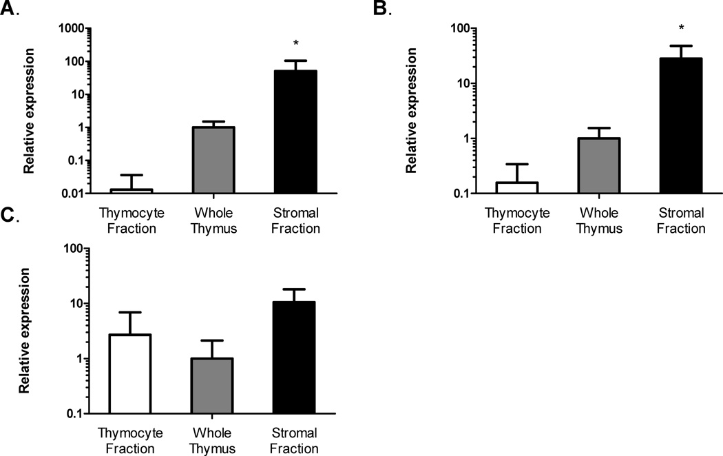
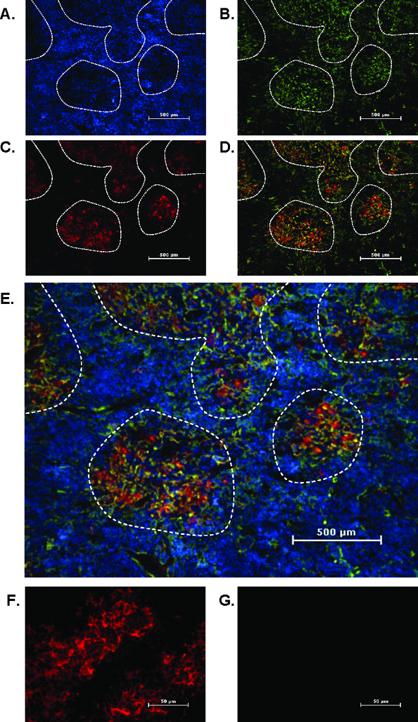
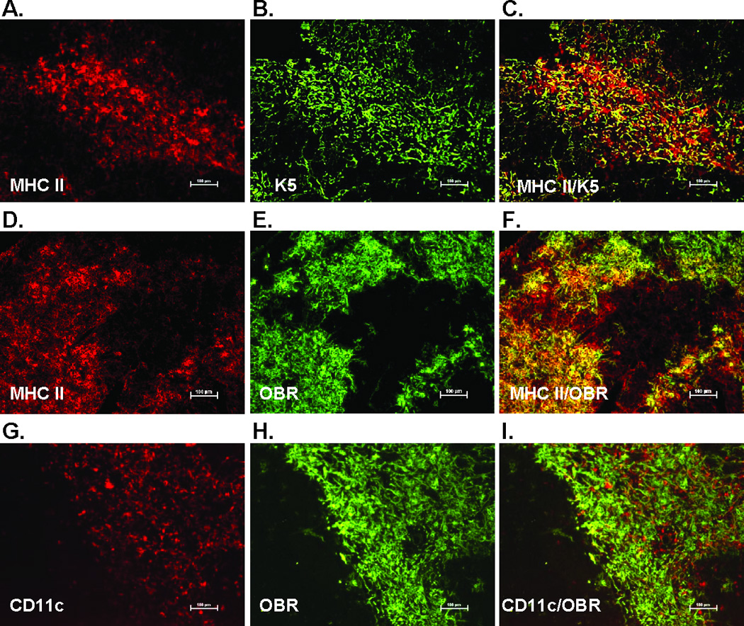
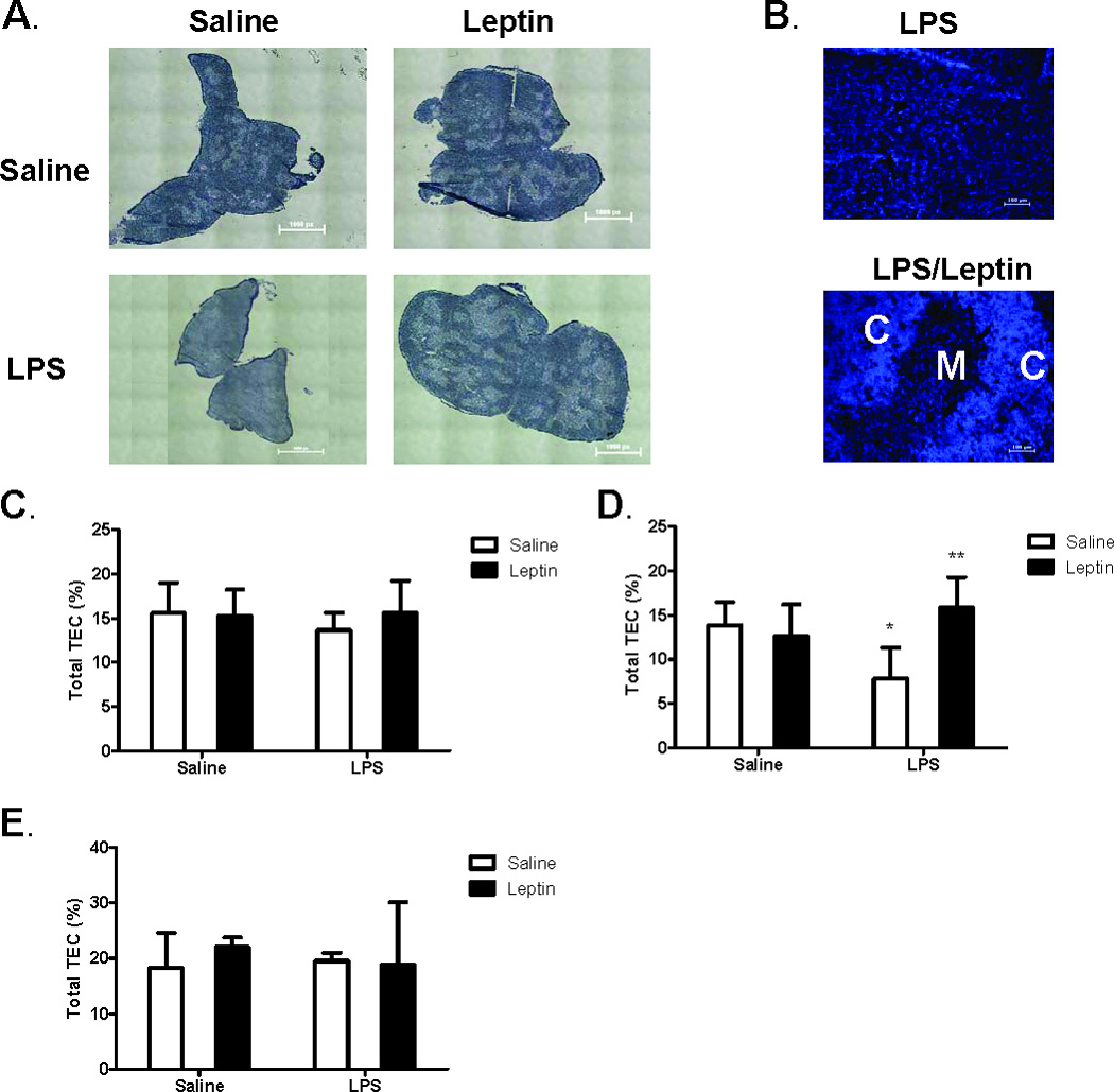
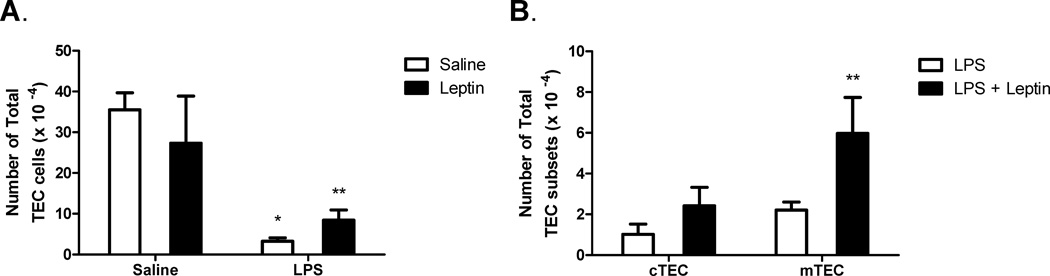
Leptin deficiency in mice results in chronic thymic atrophy, suppressed cell-mediated immunity, and decreased numbers of total lymphocytes, suggesting a key role for the metabolic hormone leptin in regulating thymopoiesis and overall immune homeostasis. Unfortunately, the thymus is highly susceptible to stress-induced acute involution. Prolonged thymus atrophy in stress situations can contribute to peripheral T cell deficiency or inhibit immune reconstitution. Little is known, however, about specific roles for leptin signaling in the thymus or the underlying mechanisms driving thymic involution or thymic recovery after acute stress. We report here that leptin receptor expression is restricted in thymus to medullary epithelial cells. Using a model of endotoxemia-induced acute thymic involution and recovery, we have demonstrated a role for supraphysiologic leptin in protection of thymic epithelial cells (TECs). We also present data in support of our hypothesis that leptin treatment decreases in vivo endotoxemia-induced apoptosis of double positive thymocytes and promotes proliferation of double negative thymocytes through a leptin receptor isoform b-specific mechanism. Furthermore, our studies have revealed that leptin treatment increases thymic expression of interleukin-7, an important soluble thymocyte growth factor produced by medullary TECs. Taken together, these studies support an intrathymic role for the metabolic hormone leptin in maintaining healthy thymic epithelium and promoting thymopoiesis, which is revealed when thymus homeostasis is perturbed by endotoxemia.


Conflict of interest statement

Disclosures

The authors have no financial conflicts of interest.

Figures

Figure 1
Figure 1. Leptin receptor (OBR) gene expression is greater in thymus stroma than thymus lymphoid compartment
Fold-change of OBRa (A), OBRb (B), and OBRc (C) steady-state mRNA isoform gene expression in thymocytes and thymic stroma fractions compared to whole thymus (Set to 1). Data presented are mean ± SD from three mice per group. *p ≤ 0.05, stromal fraction versus thymocyte fraction.
Figure 2
Figure 2. Leptin receptor (OBR) protein expression is restricted to thymic medullary epithelial cells
Frozen thymus sections from 12 week old mice were stained with an mTEC marker, cytokeratin 5 (K5) (green), and leptin receptor (pan marker, red). Nuclei were stained with DAPI (blue). Panels depict staining for DAPI (A), cytokeratin 5 (B), leptin receptor (C), leptin receptor and K5 overlay (D) and leptin receptor and K5 overlay with DAPI (E). Final magnification 40X. Areas of medulla are outlined by dashed line. Final magnification 400X shows leptin receptor (red) (F) or isotype control antibody (G). Representative section from five mice per group.
Figure 3
Figure 3. Leptin receptor (OBR) protein expression is not detectable on thymocytes
Representative flow cytometry analysis of mouse 3T3-L1 cells (positive control) (A) and isolated thymocytes from 12 week old mice (B) stained for leptin receptor (open curve) or isotype control (shaded curve). Cytospin preparation of 3T3-L1 cells (C, E) or isolated thymocytes (D, F) stained for the leptin receptor (red) and with the nuclear stain DAPI (blue). Final magnification 100X. Representative of three mice per group.
Figure 4
Figure 4. Leptin receptor (OBR) colocalized to MHC Class II+ thymic epithelial cells but not thymic dendritic cells
Frozen thymus sections from 12 week old mice were stained with the thymic epithelial cell marker K5, MHC Class II (red) cytokeratin 5 (K5)(green), dendritic cell marker CD11c (red), and leptin receptor (pan marker, green). Final magnification 100X. Representative section from three mice per group.
Figure 5
Figure 5. Leptin-mediated protection of thymic stroma during LPS challenge
Female C57BL/6 mice were administered either saline or leptin (1 µg/g body weight, IP) and simultaneously challenged with either saline or E. coli LPS (100 µg/mouse, IP). Hematoxylin and Eosin staining of representative thymus sections (A) seven days post treatment. Whole tissue stitched image, total magnification (40X). Calibration 1.61 µm equals 1.0 pixels. DAPI staining of thymocyte nuclei (B) depict distinct areas of cortex (C) and medulla (M). Total magnification (100X). Frequency of thymic epithelial cells (MHC Class II+ cells) in CD45- stromal cell population one day (C), three days (D), and seven days (E) post treatment. Data presented are mean ± SD from four mice per group. * p ≤ 0.05 compared with saline-treated controls. ** p ≤ 0.05 compared with LPS-treated controls.
Figure 6
Figure 6. Leptin-mediated protection of thymic epithelial cells during LPS challenge
Female C57BL/6 mice were administered either saline or leptin (1 µg/g body weight, IP) and simultaneously challenged with either saline or E. coli LPS (100 µg/mouse, IP). Total number of thymic epithelial cells (CD45-, MHC Class II+) three days post treatment (A). Total number of cTEC (CD45-, MHC Class II+, Ly-51+) and mTEC (CD45-, MHC Class II+, Ly-51-) cells three days post treatment (B). Data presented are mean ± SD from four mice per group. * p ≤ 0.05 compared with saline-treated controls. ** p ≤ 0.05 compared with LPS-treated controls.
Figure 7
Figure 7. Leptin-induced thymic protection is characterized by increased thymocyte proliferation and decreased thymocyte apoptosis
Female C57BL/6 mice were administered either saline or leptin (1 µg/g body weight, IP) and simultaneously challenged with either saline or E. coli LPS (100 µg/mouse, IP). Frequency of DP thymocytes (A) and frequency of DN thymocytes (B) were determined one and three days post treatment, respectively. Frequency of DN thymocytes positive for BrdU (C) was determined three days post treatment. Frequency of DP thymocytes positive for active caspase 3 was determined in C57BL/6 (D) or db/db (E) mice one day post treatment. Percent reduction of active caspase 3-positive cells in LPS challenged mice with leptin treatment in C57BL/6 and db/db mouse strains (F). Data presented are mean ± SD from five mice per group.* p ≤ 0.05 compared with saline-treated controls. ** p ≤ 0.05 compared with LPS-treated controls.

Similar articles

Cited by

References

    1. Ahima RS, Osei SY. Leptin signaling. Physiology and Behavior. 2004;81:223–241. - PubMed
    1. Bjorbaek C, Uotani S, da Silva B, Flier JS. Divergent signaling capacities of the long and short isoforms of the leptin receptor. Journal of Biological Chemistry. 1997;272:32686–32695. - PubMed
    1. Cohen P, Yang G, Yu X, Soukas AA, Wolfish CS, Friedman JM, Li C. Induction of leptin receptor expression in the liver by leptin and food deprivation. Journal of Biological Chemistry. 2005;280:10034–10039. - PubMed
    1. De Rosa V, Procaccini C, Cali G, Pirozzi G, Fontana S, Zappacosta S, La Cava A, Matarese G. A key role of leptin in the control of regulatory T cell proliferation. Immunity. 2007;26:241–255. - PubMed
    1. Erickson M, Morkowski S, Lehar S, Gillard G, Beers C, Dooley J, Rubin JS, Rudensky A, Farr AG. Regulation of thymic epithelium by keratinocyte growth factor. Blood. 2002;100:3269–3278. - PubMed

Publication types

MeSH terms