Comparative Study
. 2009 Jul 8;29(27):8639-48.
doi: 10.1523/JNEUROSCI.0898-09.2009.

Alpha-latrotoxin stimulates a novel pathway of Ca2+-dependent synaptic exocytosis independent of the classical synaptic fusion machinery

Affiliations
Comparative Study

Alpha-latrotoxin stimulates a novel pathway of Ca2+-dependent synaptic exocytosis independent of the classical synaptic fusion machinery

Ferenc Deák et al. J Neurosci. .

Abstract

Alpha-latrotoxin induces neurotransmitter release by stimulating synaptic vesicle exocytosis via two mechanisms: (1) A Ca(2+)-dependent mechanism with neurexins as receptors, in which alpha-latrotoxin acts like a Ca(2+) ionophore, and (2) a Ca(2+)-independent mechanism with CIRL/latrophilins as receptors, in which alpha-latrotoxin directly stimulates the transmitter release machinery. Here, we show that the Ca(2+)-independent release mechanism by alpha-latrotoxin requires the synaptic SNARE-proteins synaptobrevin/VAMP and SNAP-25, and, at least partly, the synaptic active-zone protein Munc13-1. In contrast, the Ca(2+)-dependent release mechanism induced by alpha-latrotoxin does not require any of these components of the classical synaptic release machinery. Nevertheless, this type of exocytotic neurotransmitter release appears to fully operate at synapses, and to stimulate exocytosis of the same synaptic vesicles that participate in physiological action potential-triggered release. Thus, synapses contain two parallel and independent pathways of Ca(2+)-triggered exocytosis, a classical, physiological pathway that operates at the active zone, and a novel reserve pathway that is recruited only when Ca(2+) floods the synaptic terminal.


Figures

Figure 1.
Figure 1.
Synaptobrevin-2 is required for Ca2+-independent but not Ca2+-dependent exocytosis induced by α-latrotoxin. A, Representative traces of mEPSCs recorded in wild-type and synaptobrevin-2 KO neurons before and after application of 0.2 nm α-latrotoxin in the presence of 2 mm Ca2+, followed by removal of Ca2+ with 4 mm EGTA, and subsequent readdition of Ca2+. Note the compressed timescale. B, Representative mEPSC traces monitored in neurons cultured from littermate wild-type (WT) and synaptobrevin-2 KO (Syb2 KO) mice. mEPSCs were obtained under control conditions in 2 mm Ca2+ (spontaneous), after addition of 0.2 nm α-latrotoxin in the same Ca2+-containing medium, and after further addition of 4 mm EGTA as indicated. The inset below illustrates the kinetics of individual synaptic events from wild-type and KO neurons. C, Summary graphs of the mean mEPSC frequency (left, note logarithmic scale), mEPSC amplitude (center), and mEPSC rise-time (right) during the three conditions shown in B: spontaneous mEPSCs before addition of α-latrotoxin (Spontaneous), after addition of α-latrotoxin in Ca2+ (α-Ltx + 2 mm Ca2+), and after further addition of 4 mm EGTA (α-Ltx + 4 mm EGTA; n = 9 synaptobrevin-2 KO, n = 14 WT neurons from 5 cultures). Data shown are means ± SEMs; calibration bars apply to all traces immediately above them. Asterisks denote significant differences between wild-type and synaptobrevin-2 KO neurons as assessed by Student's t test (*p < 0.05; **p < 0.01).
Figure 2.
Figure 2.
α-Latrotoxin stimulates synaptic exocytosis in synaptobrevin-2 KO synapses by mediating Ca2+ influx. A, B, Representative traces (A) and summary graphs of the mean frequency (B) of mEPSCs monitored in wild-type (WT) and synaptobrevin-2 KO (Syb2 KO) neurons before (Spontaneous) and after addition of N4C-mutant of α-latrotoxin in which 4 aa are inserted between the N-terminal cysteine-rich domain and the C-terminal ankyrin repeats of α-latrotoxin [0.4 nm α-LtxN4C + 2 Ca2+ (Ichtchenko et al., 1998); WT, n = 3; Syb2 KO, n = 4 from 3 cultures]. C, D, Representative traces (C) and summary graphs of the mean frequency (D) of mEPSCs monitored in WT and synaptobrevin-2 KO (Syb2 KO) neurons before (Spontaneous) and after addition of α-latrotoxin in 2 mm Ca2+, and after further addition of either 0.2 mm Cd2+ (α-Ltx + Cd2+) or 0.2 mm La3+ (α-Ltx + La3+) (WT, n = 3; Syb2 KO, n = 4 from 3 cultures). Both Cd2+ and La3+ block the Ca2+-conducting pore produced by α-latrotoxin (Chanturiya and Nikoloshina, 1994; Hurlbut et al., 1994). Data shown are means ± SEMs; calibration bars apply to all traces immediately above them. Asterisks denote significant differences between wild-type and synaptobrevin-2 KO neurons as assessed by Student's t test (*p < 0.05; **p < 0.01; ***p < 0.001).
Figure 3.
Figure 3.
The Ca2+ ionophore ionomycin potently stimulates synaptic exocytosis in synaptobrevin-2 KO neurons. A, B, Representative traces (A) and mean frequency (B) of mEPSC monitored in neurons from littermate wild-type (WT) and synaptobrevin-2 KO (Syb2 KO) mice obtained in 2 mm Ca2+ before (Spontaneous) and after addition of 0.1 mm ionomycin (Ionom. + 2 Ca2+), and after further addition of 4 mm EGTA (Ionom. + 4 EGTA; wild-type, n = 4; synaptobrevin-2 KO, n = 3 from 2 cultures). Calibration bars apply to all traces above them. Data shown are means ± SEMs; asterisks denote significant differences between wild-type and synaptobrevin-2 KO neurons as assessed by Student's t test (*p < 0.05).
Figure 4.
Figure 4.
Vesicle pools monitored by FM2-10 staining in wild-type and synaptobrevin-2 KO synapses. A, Representative fluorescence images from wild-type and synaptobrevin-2 KO hippocampal cultures. Synapses were stained with FM2-10 by α-latrotoxin stimulation (see Materials and Methods for details), and washed for 10 min. Neurons were then stimulated by application of 0.5 m sucrose, and destaining was monitored by fluorescence microscopy. Background fluorescence remaining after an exhaustive stimulation with five applications of 90 mm K+-containing Tyrode's solution was subtracted (Scale bar, 10 μm). B, Mean FM2-10 destaining time course on application of 0.5 m sucrose to neurons loaded with FM2-10 by α-latrotoxin stimulation. Destaining was measured by fluorescence microscopy in wild-type synapses (WT; n = 6, 352 synapses) and synaptobrevin-2 KO synapses (Syb2 KO; n = 5, 251 synapses from 3 cultures). C, Bar graph depicting the total amount of FM2-10 fluorescence loaded into synapses during α-latrotoxin stimulation (left; WT: 247 ± 37 vs Syb2 KO: 216 ± 33 A.U.), and destained on application of 0.5 m sucrose (right; WT: 57 ± 19 vs Syb2 KO: 34 ± 4.8 A.U.). Data shown are means ± SEMs; asterisks denote significant differences between wild-type and synaptobrevin-2 KO neurons as assessed by Student's t test (*p < 0.05). Quantitations in B and C were performed after background subtraction as described in A.
Figure 5.
Figure 5.
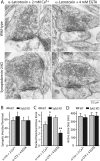
α-Latrotoxin treatment does not impair synaptic terminal integrity. A, Representative electron micrographs of wild-type and synaptobrevin-2 KO synapses after a 10 min application of 0.2 nm α-latrotoxin in Tyrode's solution containing either 2 mm Ca2+, followed by a change to Tyrode's solution with the same Ca2+ concentration, or 4 mm EGTA. Cells were fixed after 20 min in these solutions. Calibration bar applies to all images. B–D, Bar graphs depicting the mean synaptic vesicle number per nerve terminal (B), docked synaptic vesicle number per active zone (C), and size of the active zone (D; n = 11–15 synapses per condition). Data shown are means ± SEMs. Asterisks denote significant differences between wild-type and synaptobrevin-2 KO neurons as assessed by Student's t test (**p < 0.01). Other abbreviations are as defined in the legend to Figure 1.
Figure 6.
Figure 6.
Role of the SNARE-protein SNAP-25 in the active-zone protein Munc13-1 in Ca2+-dependent and Ca2+-independent α-latrotoxin-triggered release. A, B, Representative traces (A) and summary graphs of the mean frequency (B) of mEPSCs monitored in wild-type (WT) and SNAP-25 KO neurons. mEPSCs were obtained under control conditions in 2 mm Ca2+ (Spontaneous), after addition of 0.2 nm α-latrotoxin in Ca2+ (α-Ltx + 2 mm Ca2+), and after further addition of 4 mm EGTA (α-Ltx + 4 mm EGTA; n = 3 for both WT and SNAP25 KO from 3 cultures). Note the logarithmic ordinate. C, D, Same as A and B, respectively, except that wild-type and Munc13-1 KO neurons were compared (WT, n = 4; Munc13-1 KO, n = 5 from 3 cultures). Data shown are means ± SEMs; calibration bars apply to all traces immediately above them. Asterisks denote significant differences between wild-type and synaptobrevin-2 KO neurons as assessed by Student's t test (*p < 0.05; **p < 0.01; ***p < 0.001).
Figure 7.
Figure 7.
Synaptobrevin-2 with a 12-residue insertion between the SNARE motif and the membrane is unable to mediate Ca2+-independent α-latrotoxin-triggered release in synaptobrevin-2 KO neurons. A, Schematic illustration of eCFP-tagged synaptobrevin-2 and its mutant version with a 12-residue insertion between the SNARE motif and transmembrane region. B, C, Representative traces of mEPSCs (A) and summary graphs of the mean mEPSC frequency (B) monitored in wild-type (WT) and synaptobrevin-2 KO neurons that were infected with lentivirus expressing wild-type synaptobrevin-2 (Syb KO + Syb) or mutant synaptobrevin 2 with the 12-residue insertion (Syb KO +Syb12ins). mEPSCs were monitored under control conditions in 2 mm Ca2+ (Spontaneous), after addition of 0.2 nm α-latrotoxin in Ca2+ (α-Ltx + 2 mm Ca2+), and after further addition of 4 mm EGTA (α-Ltx + 4 mm EGTA; WT, n = 13; Syb KO + Syb12ins, n = 6 from 5 cultures). Data shown are means ± SEMs; calibration bars apply to all traces immediately above them. Asterisks denote significant differences between wild-type and synaptobrevin-2 KO neurons as assessed by Student's t test (***p < 0.001).

Similar articles

Cited by

References

    1. Ádám-Vizi V, Déri Z, Bors P, Tretter L. Lack of involvement of [Ca2+]i in the external Ca2+-independent release of acetylcholine evoked by veratridine, ouabain and alpha-latrotoxin: possible role of [Na+]i . J Physiol Paris. 1993;87:43–50. - PubMed
    1. Augustin I, Rosenmund C, Südhof TC, Brose N. Munc13-1 is essential for fusion competence of glutamatergic synaptic vesicles. Nature. 1999;400:457–461. - PubMed
    1. Bronk P, Deák F, Wilson MC, Liu X, Südhof TC, Kavalali ET. Differential effects of SNAP-25 deletion on Ca2+-dependent and Ca2+-independent neurotransmission. J Neurophysiol. 2007;98:794–806. - PubMed
    1. Brose N, Hofmann K, Hata Y, Südhof TC. Mammalian homologues of Caenorhabditis elegans unc-13 gene define novel family of C2-domain proteins. J Biol Chem. 1995;270:25273–25280. - PubMed
    1. Capogna M, Gähwiler BH, Thompson SM. Calcium-independent actions of α-latrotoxin on spontaneous and evoked synaptic transmission in the hippocampus. J Neurophysiol. 1996;76:3149–3158. - PubMed

Publication types

MeSH terms

LinkOut - more resources