Comparative Study
. 2009 Jul 8;29(27):8649-54.
doi: 10.1523/JNEUROSCI.1864-09.2009.

Reassessment of corticospinal tract regeneration in Nogo-deficient mice

Affiliations
Comparative Study

Reassessment of corticospinal tract regeneration in Nogo-deficient mice

Jae K Lee et al. J Neurosci. .

Abstract

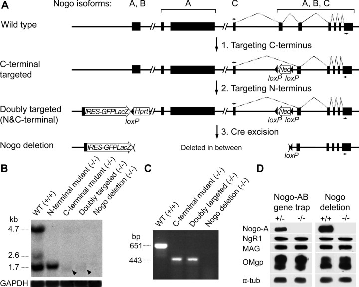
The myelin-derived neurite growth inhibitor Nogo has been proposed to play a major role in blocking axon regeneration in the CNS after injuries. However, past studies have produced mixed results regarding the regenerative phenotype of various Nogo-deficient mouse lines after experimental spinal cord injury. Two lines did not display enhanced corticospinal tract (CST) regeneration, and one displayed modest regeneration. A fourth line, a Nogo-A,B gene-trap mutant, was instead reported to exhibit extensive CST regeneration, but the results were later found to be inadvertently confounded with an axon labeling artifact. Of the four Nogo mutant lines studied so far, three continue to express some isoform(s) of Nogo, leaving open the question whether any remaining Nogo protein contributes to the modest regenerative phenotype reported in some. The remaining Nogo mutant line studied was confounded by the unexplained rescue of embryonic lethality associated with this mutation. To gain a better understanding of the contribution of Nogo as an inhibitor of regeneration of CNS axons, and particularly CST axons, we reanalyzed the Nogo-A,B gene-trap mutant line and analyzed a novel, fully viable Nogo deletion mutant line that is null for all known isoforms of Nogo. Our analyses failed to reveal any enhanced CST regeneration after experimental spinal cord injury in either line. These results indicate that Nogo alone does not account for lack of CST regeneration and have implications for current therapeutic development for spinal cord injury in humans by targeting Nogo.


Figures

Figure 1.
Figure 1.
Analysis of Nogo-A,B gene-trap mutant mice for CST regeneration and locomotor recovery. A, B, Representative sagittal spinal cord sections showing the labeled main CST tract approaching but not passing through the injury site (arrow). Rostral is to the left. Het, Heterozygous; KO, knock-out (mutant). Scale bar, 500 μm. C, Quantification of CST axons along the rostrocaudal axis on sagittal sections did not reveal any significant differences between the two genotypes. D, Locomotor recovery as assessed by a modified BBB open-field score did not show any significant differences between the two genotypes.
Figure 2.
Figure 2.
Generation and molecular characterization of the Nogo deletion mutant. A, The strategy to obtain the Nogo deletion allele via two consecutive gene targeting steps followed by Cre-mediated excision. A, B, C above the exons (black bars) refer to Nogo isoforms. Dotted lines, Splicing pattern for the Nogo-C transcript; Neo, neomycin resistance gene; Hprt, hypoxanthine phosphoribosyltransferase gene; IRES, internal ribosomal entry site; GFPLacZ, green fluorescent protein and β-galactosidase fusion gene; small arrows, primers used to analyze the Nogo-C transcript by RT-PCR. Two Frt sites flanking Hprt are omitted for simplicity. B, Northern blot with a C-terminal common probe. Nogo-A,B,C transcripts run at 4.7, 2.6, and 1.7 kb, respectively. Arrowheads indicate a slightly shorter, mutated form of Nogo-C transcript. WT, Wild type. C, RT-PCR to confirm the absence of Nogo-C transcript in the Nogo deletion mutant. D, Western blot analysis on Nogo-A, NgR1, MAG, and OMgp in Nogo-A,B gene-trap and Nogo deletion mutants. α-tub, α-Tubulin.
Figure 3.
Figure 3.
Analysis of Nogo deletion mutant mice for CST regeneration. A, B, Representative sagittal spinal cord sections showing the labeled main CST tract approaching but not passing through the injury site (arrow). ND, Nogo deletion; WT, wild type; KO, knock-out. Rostral is to the left. Scale bar, 500 μm. C, Quantification of CST axons caudal to the injury site did not indicate any significant differences between the two genotypes, but wild-type mice showed significantly more axons at two distance points rostral to the injury site. *p < 0.05 two-way repeated measures ANOVA with Bonferroni's post hoc test.

References

    1. Basso DM, Beattie MS, Bresnahan JC. A sensitive and reliable locomotor rating scale for open field testing in rats. J Neurotrauma. 1995;12:1–21. - PubMed
    1. Brösamle C, Huber AB, Fiedler M, Skerra A, Schwab ME. Regeneration of lesioned corticospinal tract fibers in the adult rat induced by a recombinant, humanized IN-1 antibody fragment. J Neurosci. 2000;20:8061–8068. - PMC - PubMed
    1. Cafferty WB, Kim JE, Lee JK, Strittmatter SM. Response to correspondence: Kim et al., “axon regeneration in young adult mice lacking Nogo-A/B.” Neuron 38, 187–199. Neuron. 2007;54:195–199. - PMC - PubMed
    1. Cafferty WB, McGee AW, Strittmatter SM. Axonal growth therapeutics: regeneration or sprouting or plasticity? Trends Neurosci. 2008;31:215–220. - PMC - PubMed
    1. Caroni P, Schwab ME. Two membrane protein fractions from rat central myelin with inhibitory properties for neurite growth and fibroblast spreading. J Cell Biol. 1988a;106:1281–1288. - PMC - PubMed

Publication types